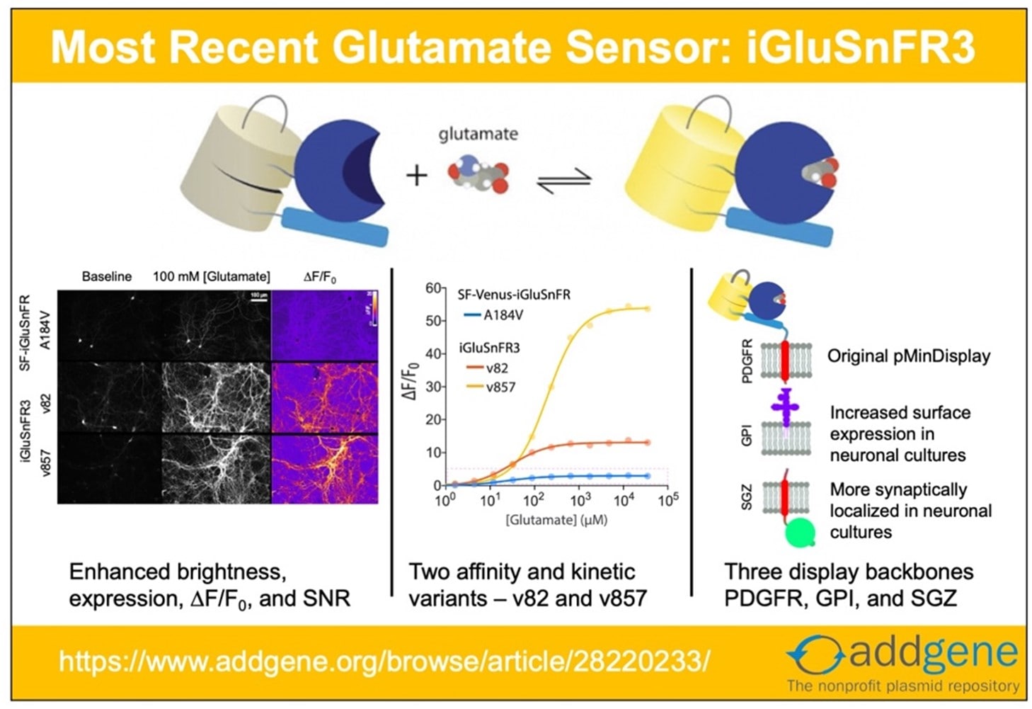
This post was originally written by Tyler Ford in 2018. It was updated by guest blogger Abhi Aggarwal in 2022. Recent updates to iGluSnFR and SF-iGluSnFR have made it clear that it’s time to update our iGluSnFR post! Here, we look at the origins of the system and explore ...
Many neuroscience experiments that require gene expression in a specific cell type rely on transgenic models that express recombinases like Cre or Flp in their cells of interest and recombinase-dependent AAV vectors for selective transgene expression. While this is a powerful ...
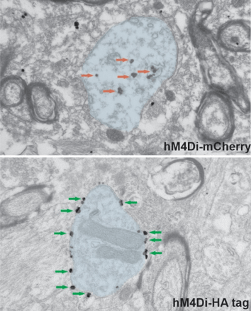
This post was contributed by Adriana Galvan, an associate professor at Emory University School of Medicine. Optogenetics and chemogenetics are powerful tools to modulate the activity of neurons and other brain cells. Since the opsins or chemogenetic receptors used in these ...
To deliver genes using lentiviral vectors, you need an envelope protein on the virus’s surface and a corresponding receptor in the host cell. Some of these envelope-receptor pairings are broad, allowing delivery into many cell types, while others are specific, allowing delivery ...
Your next cool experiment requires some AAV. Where do you start? Plasmids of course! You just need three plasmids to start making the AAVs you need for your experiment: the packaging plasmid which contains the AAV structural and packaging genes, the adenoviral helper plasmid ...
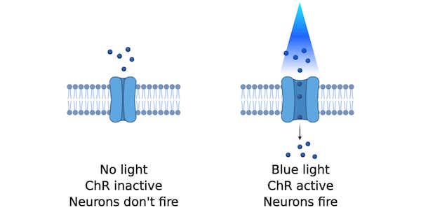
Optogenetics is a neuroscience method that lets you fire neurons with the flick of a light switch. Neurons are not typically persuaded to fire when light is shined on them, but the expression of light-gated ion channels such as channelrhodopsins (ChRs) makes them ...
This post was contributed by guest blogger, Kaustubh Kishor Jadhav, a Research Assistant at MGMs Institute of Biosciences and Technology. If you are reading this article then you probably suspect mycoplasma contamination in your cell culture or you are about to begin a new cell ...
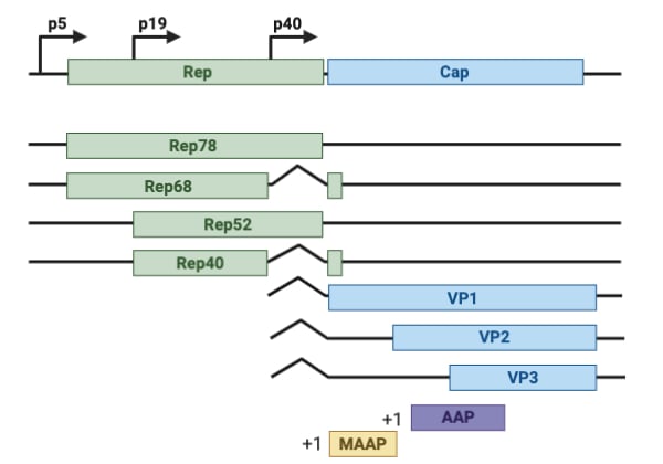
Adeno-associated virus (AAV) has many features which make it a great viral vector, but its packaging capacity is limited to ~4.7kb, or roughly half the packaging limits of lentiviral and adenoviral vectors. While many transgene will fit within this limit, some like prime ...


.png)