Master plasmid fundamentals, CRISPR techniques, AAV serotype selection, and antibody applications. Written by scientists, for scientists.
Subscribe
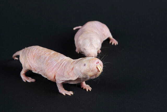
Modeling is not just a job for humans — it is also a job for many different organisms. And like human modeling, biological modeling turns out to be surprisingly glamorous and diverse. But what comes to mind when you picture a “model organism”? A mouse? A pesky fruit fly? ...
For many PhDs, the transition from graduate school or a postdoc into the next career step can feel like running an experiment without a clear protocol. You may have spent years optimizing conditions, troubleshooting assays, and becoming deeply familiar with one narrow scientific ...
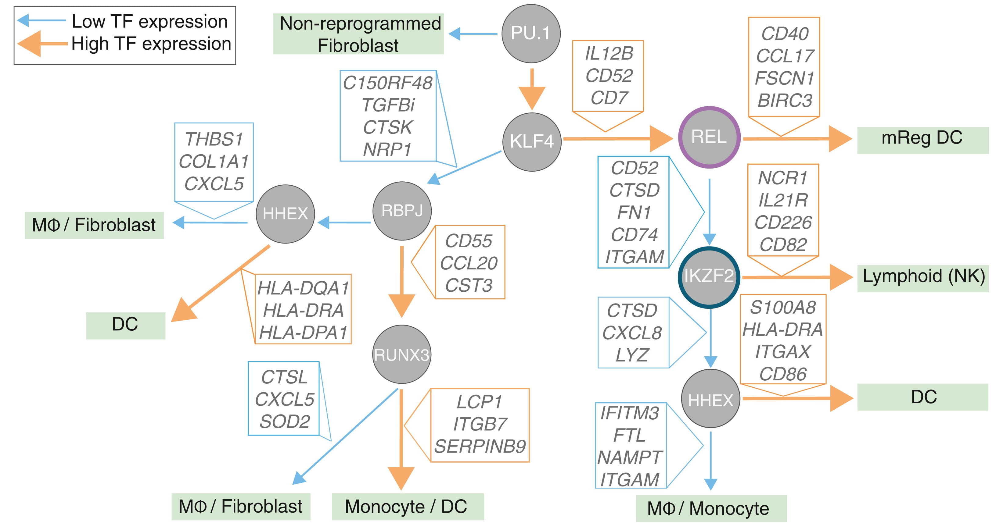
Guiding a cell through differentiation to its eventual fate requires a precise recipe of molecular signals, with transcription factors (TFs) as the key ingredients. With the right combination of TFs, scientists can also reprogram cell identity in the lab. In immune cells, such ...
iGEM isn’t any old science fair. “The world’s biggest synthetic biology competition” brings together hundreds of student teams to build projects and solve problems using molecular biology.
Every few months we highlight some of the new plasmids, antibodies, viral preps, and other materials in the repository through our Hot Plasmids articles. This month, we’ve got some new biosensors for neuroscience research, popular plasmids for a common lab enzyme, and more!
Check us out! This week Addgene rolled out our new brand identity, and we’re feeling sharp. We’re so excited to show you what we’ve been working on.
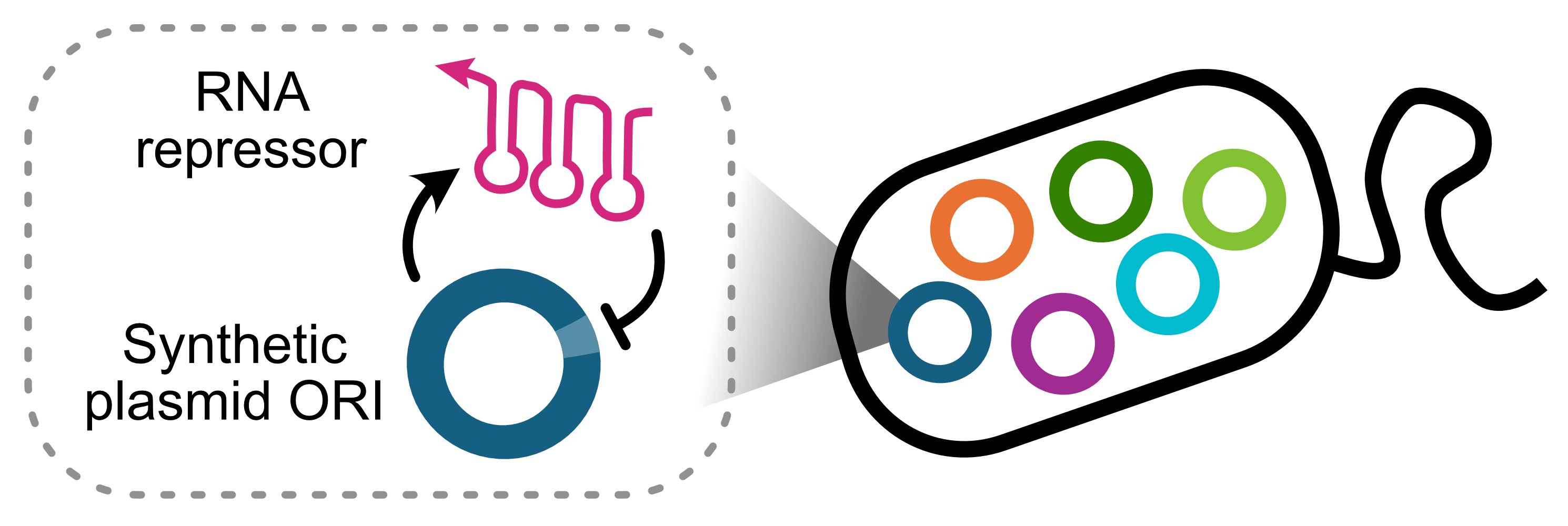
By Baiyang Liu and James Chappell, Rice University. For decades, we’ve been designing experiments around two major limitations of plasmids: copy number and incompatibility. While functional, such workarounds are clunky. To address this, we created a synthetic origin of ...
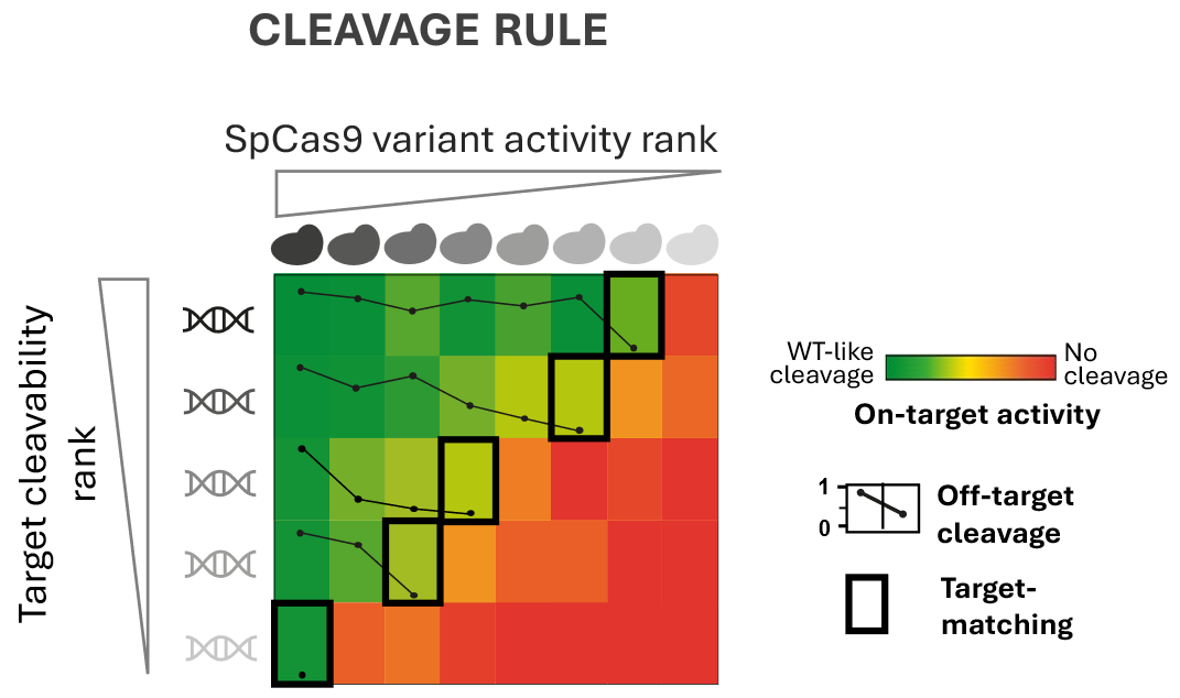
ByBalázs Csoma, Hungarian Research Network CRISPR nucleases are remarkably precise molecular tools for cutting DNA. But “remarkably precise” does not mean “perfectly specific.” In reality, CRISPR nucleases occasionally make mistakes and cleave DNA sequences that only resemble ...