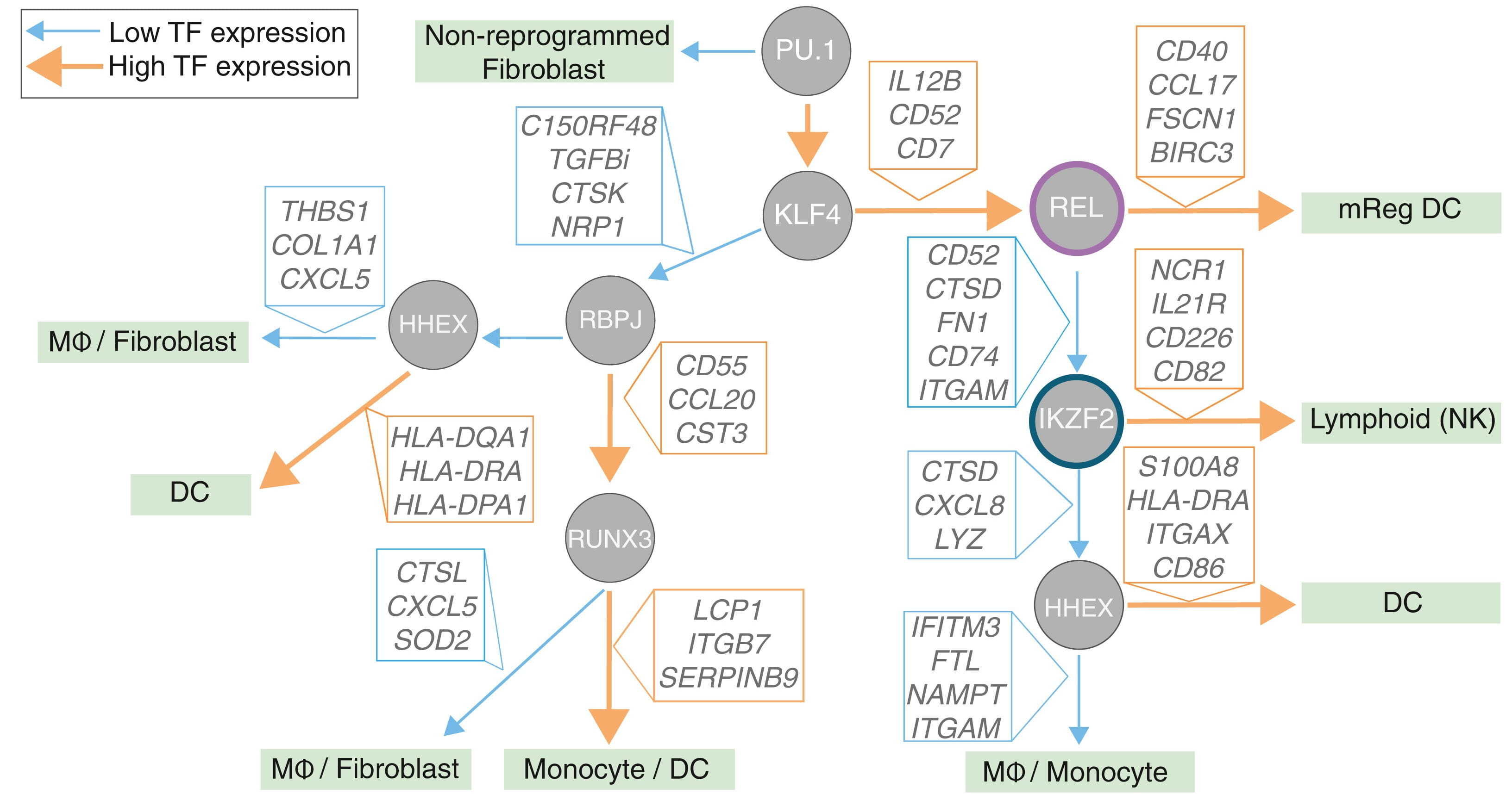
Guiding a cell through differentiation to its eventual fate requires a precise recipe of molecular signals, with transcription factors (TFs) as the key ingredients. With the right combination of TFs, scientists can also reprogram cell identity in the lab. In immune cells, such ...
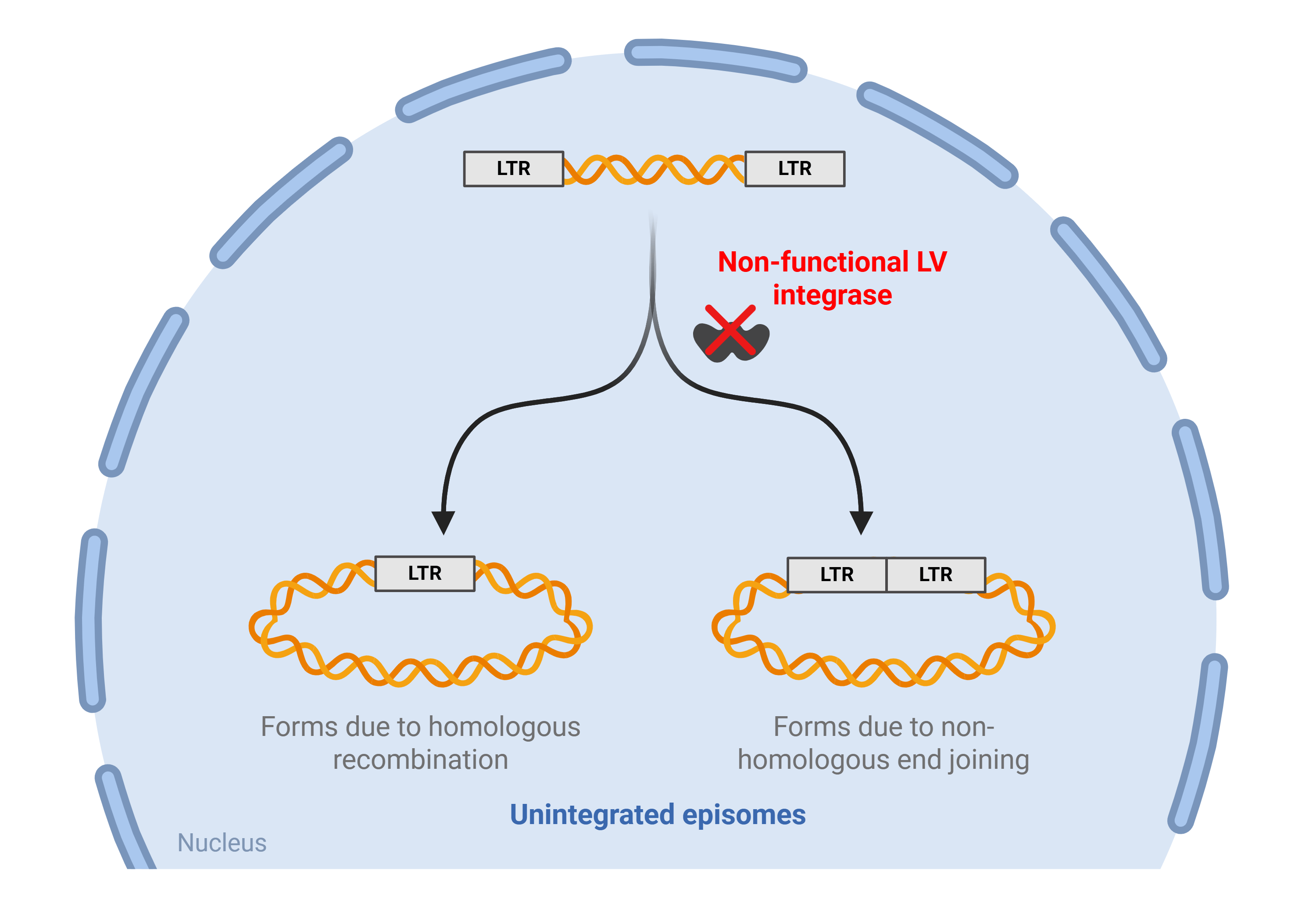
Lentiviral vectors have been a staple in molecular biology for over three decades. Widely used across various research applications, they have become indispensable tools for manipulating cells and organisms. They can be used for a variety of research purposes
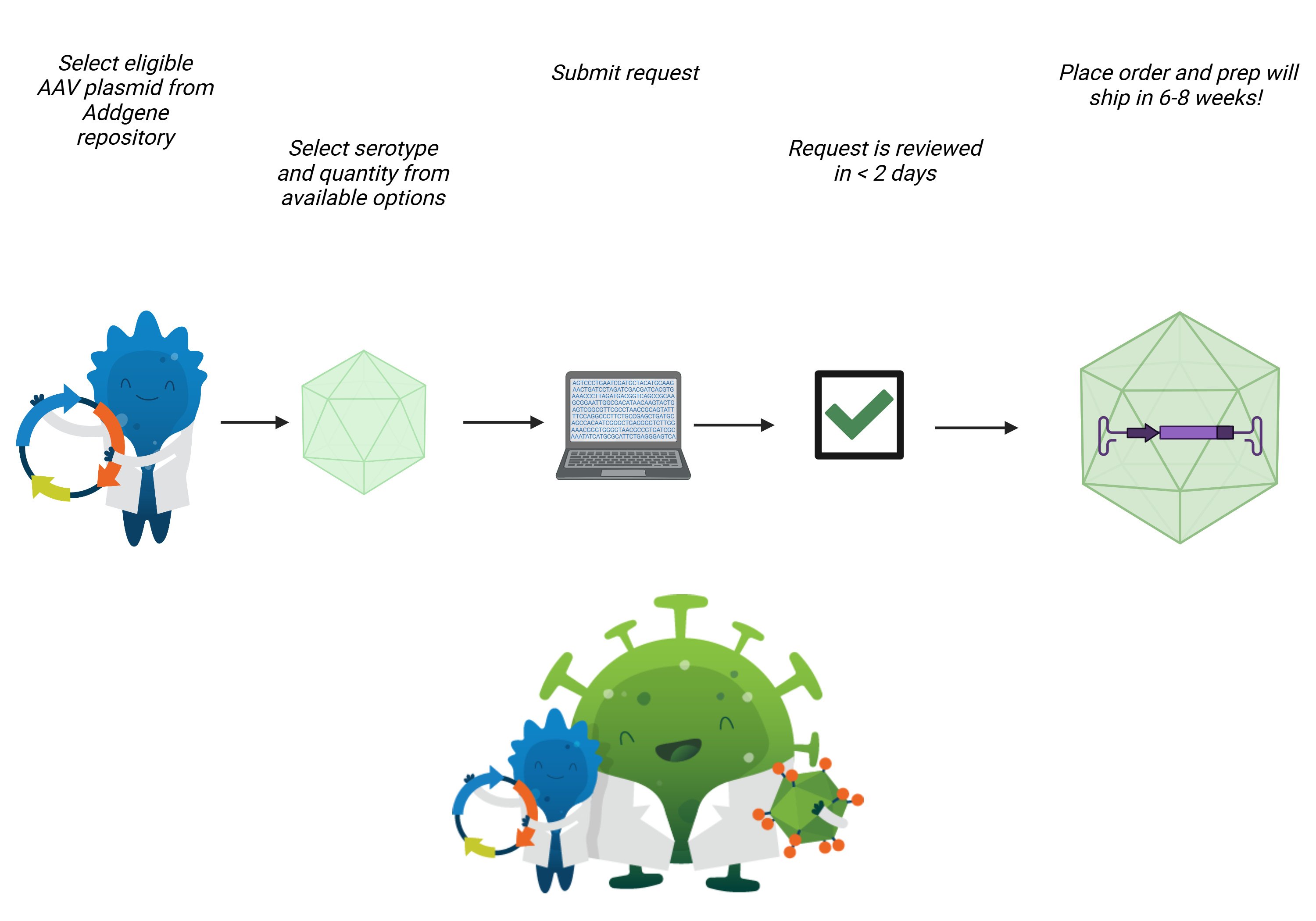
Addgene’s viral vector services allow scientists to conduct experiments faster, skipping the preparation steps of production, purification, and titering. Our newest viral vector service, AAV Packaged on Request has seen a fantastic response from the research community. Customers ...
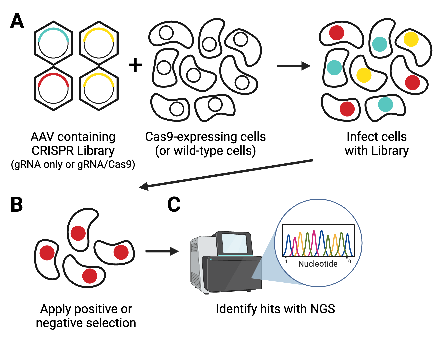
Forward genetics screens are a valuable part of the molecular biology toolbox to identify new target genes for drug discovery or to understand the intricacies of molecular pathways. These screens have gotten larger, easier, and more comprehensive thanks to the consistent ...
Bundling up for winter? The weather outside might be frightful, but browsing the 49 new preps available in the Addgene viral vector repository with a nice cup of hot chocolate is absolutely delightful. Grab a hot beverage and a blanket, then scroll on for some wintery delights!
AAV Packaged on Request, an expansion of our viral vectors service, has officially launched! As of today, you can request any of over 3,000 eligible AAV cargo plasmids in the Addgene repository to be packaged in your choice of five different serotypes.
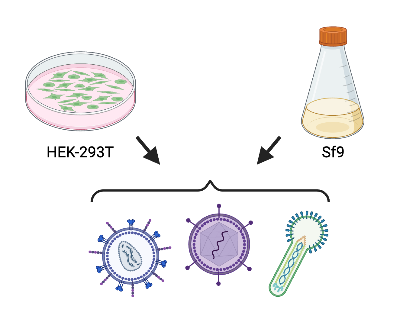
Pre-made viruses have become increasingly accessible and are useful for saving time and avoiding potentially costly set-ups. However, there are many cases where the specific viral particles you need are not available, or the cost of custom viruses are too high for your budget. ...
We’re very excited to announce that we are expanding our viral vector service to include a Packaged on Request option. Through this expansion, scientists will be able to request an AAV composed of an AAV plasmid in the Addgene repository and their choice of five different ...


.png)