This post was contributed by guest blogger, Kaustubh Kishor Jadhav, a Research Assistant at MGMs Institute of Biosciences and Technology.
If you are reading this article then you probably suspect mycoplasma contamination in your cell culture or you are about to begin a new cell culture project. If mycoplasmas are present in your lab, don’t be surprised. They are present in most of the cell culture facilities, tissue culture labs and every cell culturist has to deal with this problem. It is estimated that mycoplasma is responsible for up to 60% of the cell culture contamination (Uphoff, 2002).
Mycoplasmas are considered to be one of the simplest and smallest bacteria. The absence of a rigid cell wall makes them resistant to antibiotics and antibacterial drugs like penicillin and streptomycin. Mycoplasma can pass through filtration methods because of its ability to change shape and the absence of a rigid cell wall. Here, I will cover some of the best ways to tackle mycoplasma contamination before they enter your cell culture and what to do if you encounter mycoplasma contamination.
Causes of mycoplasma contamination
The mycoplasmas enter the cell culture through various sources that are difficult to trace. These include the laboratory personnel, the serum, the cell culture media, water baths, incubators, etc. Contamination by humans accounts for the largest source among those mentioned above. The contaminants can spread through dirty clothing, lab wears, human speech near the laminar airflow, the human scalp, sneezing, coughing, etc. Also, a constant in-flow of individuals wherever the cell cultures are kept will increase the risk of contamination.
Mycoplasmas can spread from these sources through cross-contamination and due to poor lab techniques. These include reusing pipettes for multiple cell lines rather than using disposable ones or reusing gloves. The serum is also a source of mycoplasma contamination. Due to its turbid nature, contamination is difficult to be detected in the serum that is most commonly used for every cell culture. Reusing the same bottle of serum again and again for each subculture can enhance the growth of mycoplasmas. The serum is nutrient-rich and also the best source for mycoplasma proliferation. Plus, it is added to the media after autoclaving hence, there is no assurance of the serum being contamination free.
The aerosols that are generated (while talking or pipetting) during working in the laminar airflow are also among the leading causes of contamination. These aerosols generated will enter the culture medium during subculturing processes or from the air if the cell line remains exposed for a longer time. The aerosols are invisible to naked eyes and hence cannot be detected until it leads to contamination.
Another source of the problem is the media, used for subculturing, which is available in both liquid and powdered forms. Due to improper handling or poor lab techniques, mycoplasma can spread through this form of powdered media. Filtration does not ensure complete sterilization as mycoplasma can even escape 0.2-micron filters.
-min.jpg?width=600&name=IMG20190930122344%20(2)-min.jpg)
| Figure 1: Contamination! Tissue culture media often have indicators included in the media that turns yellow from lowered pH due to microbial contamination. Photo by Kaustubh Kishor Jadhav. |
Mycoplasmas can remain in the dry state for a longer time. When they come in contact with a nutrient source, they start proliferating shortly. Once they are present in the laboratory environment, they are difficult to eradicate. You can suppress the growth of mycoplasma but cannot eradicate it completely. When mycoplasmas are seen in the culture, it is a good option to discard the flasks (unless the source of culture is irreplaceable or highly expensive).
Detecting mycoplasma contamination
Detection of mycoplasma by naked eyes or optical microscope can be very difficult, so how do you know it’s there? For this task, you can use PCR based detection, DNA staining, fluorescent tagging, agar plating, etc. Be wise to choose any of the techniques listed below and always use a second assay to be 100% sure. Before testing for mycoplasma, the cells should be in continuous culture for at least two weeks to give time for low levels of contaminations to grow. For testing, the medium should not have been changed for at least two or three days before sampling (Uphoff and Drexler, 2011).
- Culturing on agar plates/broth is considered the ‘gold standard’ for detecting mycoplasmas. In this method, the supernatant from the cell culture is added to the liquid or semi-solid medium (containing nutrients essential for mycoplasma growth). Infected cell culture will show growth both in the liquid broth and on the agar plates. Mycoplasma colonies are easily visible on the agar plates. This method is capable of detecting most of the mycoplasma species, apart from a few of them.
- Another method used for mycoplasma detection is using PCR. In this technique, PCR primers specific to the 16s RNA genes of various commonly infecting mycoplasmas are used. A broad range of primers encompasses almost all the infecting mycoplasma. The primers used in the process are either species/gene-specific or universal. This method is quick, efficient, reliable and cost-effective (Sung, 2006).
- The sensor cell-based method uses specialized cells that can detect the presence of mycoplasmas in the culture. Once these cells detect mycoplasmas, they trigger a series of chemical reactions and this induces a noticeable color change. This method has high sensitivity but low specificity (Degeling, 2012).
- Bioluminescence assay kits are available from different biotech companies which are enzyme-based mycoplasma detection kits. These kits detect mycoplasma enzymes that are not synthesized by eukaryotic cells. First, the kit components rupture the mycoplasma and then specific enzymes are used which trigger cellular mechanisms that generate luminescence. This can then be detected by a luminometer.
- Non-specific DNA stains can be added in the infected culture medium to detect mycoplasma. When observed under a fluorescent microscope, the mycoplasma DNA appears in the form of small clusters, apart from the cellular DNA.
- Fluorescent DNA staining is another DNA staining alternative. This method uses DNA stains like DAPI and Hoechst 33258 to stain all DNA. Some amount of expertise is required in this process because result interpretation might be difficult. The low density of mycoplasmas, contamination by other bacteria or extracellular fluorescent signals produced by these bacteria can hinder correct result interpretation. Also, the signals from the nuclear area make the detection of mycoplasmas difficult (Ligasová, 2019).
Prevention of mycoplasma contamination
The best and the most important ways to avoid mycoplasma are by providing proper training and guidance to the people working in the lab facility. This can be done by training eligible staff for the cell culture process and hence eliminate one of the causes of contamination. If possible, a separate lab should be made available for cell culturing and maintenance of the cell lines. Providing such an environment for the lab facility will not only reduce mycoplasmas but also avoid other bacterial and fungal contamination.
Proper PPE
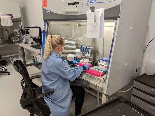
Since humans are the major source of contamination, the prevention steps start from you. If you are careful enough to provide a sterile environment for work, it will soon become a habit before you even realize it. Here are some things to do:
- Wear a clean lab coat and mask while handling cell lines.
- Avoid discussion with other lab members, sneezing or coughing in the lab. The human mouth contains a wide variety of mycoplasmas and these can enter the cell culture when you talk, laugh, sneeze or cough.
- Wear gloves while working and discard them after use. Human contact can spread contamination when you are working without gloves or masks. Make sure to change the gloves and masks regularly instead of keeping them in the pockets of your lab coats.
- Have separate footwear for the lab to avoid bringing in environmental contaminants.
 |
| Figure 2: An Addgenie works in the tissue culture hood while wearing PPE. Photo by Kate Harten DeMaio. |
Cell lines and reagents
- Examine your cell line stock for mycoplasma contamination before use.
- Make sure there is no contamination present in the serum to be used for subculturing. Avoid using expired products or reagents that have been stored for a longer time.
- Use fresh media as much as possible and the ones that are tightly packed with seals.
- Use disposable pipettes to avoid cross-contamination.
- Make sure you keep all the required equipment and reagents inside the laminar airflow before and while you are working.
- Perform routine checks for mycoplasma contamination, before using the cell lines for assay or analytical purposes.
- Use 0.1-micron filters as are they are a better way of avoiding mycoplasma, rather than using 0.2-micron filters, for media filtration. There is no 100% assurance that this process can avoid contamination but it’s a very good practice for prevention.
- Prepare a small quantity of media stock that would be used for the experiment, preventing contamination of the rest.
- Avoid long exposure of the cell culture to air and make sure to tighten the flask cap before placing them in the incubators. This will also solve the problem of contamination through aerosols.
- Perform culturing without the use of antibiotics since the use of antibiotics can allow mycoplasma to develop resistance to the antibiotics. Antibiotics must only be used to remove mycoplasma and not for preventing mycoplasma growth. Also, prolonged use of a particular antibiotic can induce resistance for other bacteria apart from mycoplasma.
- Always have a backup in the form of cryopreserved vials of the cell lines even though the viability of cryopreserved cells decreases with every subculture.
Equipment
- Fumigate the laminar airflow and the lab facility periodically.
- The CO2 incubators, used for maintaining cell lines, must not be overcrowded (filled no more than 60% of its capacity) which can cause mycoplasma infection from one cell line to another.
- Multiple cell lines should not be stored in a single incubator since it increases the risk of cross-contamination.
- CO2 incubators should be cleaned regularly and dampness of any kind should be avoided. Distilled water provided to maintain the humidity should be changed regularly as mycoplasma, bacteria, and fungus can reside in these containers.
- Clean up any media or culture spilled immediately.
- Water baths, liquid nitrogen containers, reagent bottles and other sources of infection must be cleaned regularly and checked for contamination periodically.
For more tips, check out Addgene's video Getting Started with Tissue Culture below!
Mycoplasma removal after detection
So you found mycoplasma contamination, what do you do next? The best way is to discard the infected cell culture flasks. In cases where the culture is irreplaceable or very expensive, there are some ways to rescue the cultures.
Elimination is a very time-consuming process and increases the risk of secondary contamination to other healthy cell lines. Mycoplasma Removal Agents (MRA) are derivatives of the quinolone family of antibiotics and are broad-spectrum antibiotic agents. These agents can be used to wash the cell cultures. The treatment can take a few weeks to a couple of months depending on the severity of the infection. Plasmocin is a widely used drug that can clear most of the mycoplasmas present in the culture media. Also, drugs like BM Cyclin, fluoroquinolone ciprofloxacin, ciprobay, zagam, baytril, tetracycline, etc. are available for mycoplasma removal from the infected culture.
MRAs are non-toxic, effective in the removal of most of the mycoplasma contamination, easy to use, and effective for wide range of mycoplasma. These methods cannot guarantee complete removal of mycoplasma but, can get rid of most of mycoplasma contamination and are the last alternative. There are some drawbacks while considering this treatment as the duration of treatment can affect the cell growth and viability. If mycoplasma has made permanent damage to the cell culture it is irreparable. Some cells may die due to longer incubation periods and affect the total cell viability.
Conclusion
To sum up, it is always important to prevent contamination rather than eradicating it later. Don’t be scared when mycoplasma infects your cell cultures because you are not the first person to face this kind of problem. Educate others when you can and help them understand the importance of preventing contamination. Be aware and cautious rather than learning it the hard way!
 Many thanks to guest blogger Kaustubh Kishor Jadhav!
Kaustubh Kishor Jadhav, MSc is a Research Assistant at the MGMs Institute of Biosciences and Technology in India. Jadhav has a Master's degree in Molecular Biology (University of Milan, Italy) and a Bachelor's degree in Biotechnology.
References
Degeling MH, Maguire CA, Bovenberg MSS, Tannous BA (2012) Sensitive Assay for Mycoplasma Detection in Mammalian Cell Culture. Anal Chem 84:4227–4232 . https://doi.org/10.1021/ac2033112
Ligasová A, Vydržalová M, Buriánová R, Brůčková L, Večeřová R, Janošťáková A, Koberna K (2019) A New Sensitive Method for the Detection of Mycoplasmas Using Fluorescence Microscopy. Cells 8:1510 . https://doi.org/10.3390/cells8121510
Sung H, Kang SH, Bae YJ, Hong JT, Chung YB, Lee CK, Song S (2006) PCR-based detection of mycoplasma species. J Microbiol .44: 42–49
Uphoff Cc, Drexler Hg (2002) Comparative Antibiotic Eradication of Mycoplasma Infections from Continuous Cell Lines. In Vitro Cell Dev Biol Anim 38:86 . https://doi.org/10.1290/1071-2690(2002)038<0086:caeomi>2.0.co;2
Uphoff CC, Drexler HG (2011) Detecting Mycoplasma Contamination in Cell Cultures by Polymerase Chain Reaction. In: Methods in Molecular Biology. Humana Press, pp 93–103
Additional resources on the Addgene blog
- Browse Addgene's viral vectors articles
- Find tips and tricks for working with viral vectors
Resources on Addgene.org
- Check out Addgene's written and video protocols


Leave a Comment