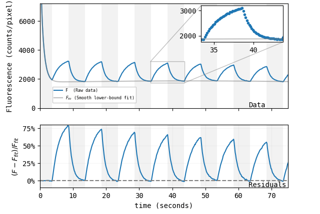
Lots of scientists work to invent novel techniques or to engineer improved tools for familiar applications. But how does one invent a tool for applications that don’t even exist yet? Andrew York, Maria Ingaramo, and their team at Calico Life Sciences recently set out to do just ...
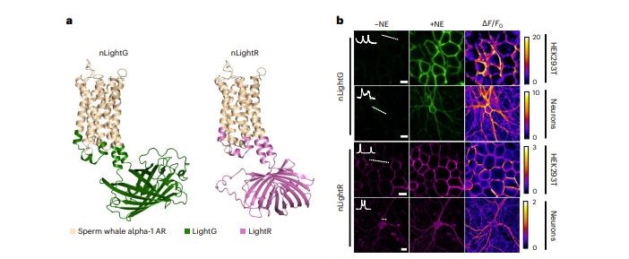
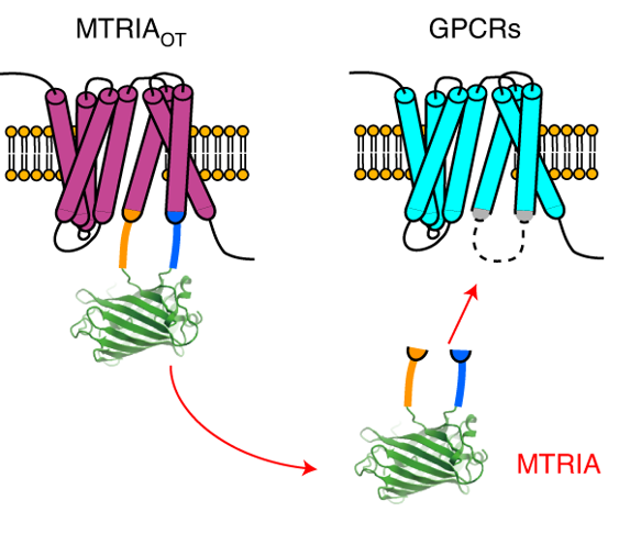
Neuromodulators like dopamine and norepinephrine have important functions in the brain but have been difficult to study without biosensors to directly visualize their activity. In 2019, the first generation of norepinephrine sensors was developed, named GRABNE, which helped ...
Here at Addgene’s headquarters, the skies are getting dark and wintry with the end of year approaching. We’re always looking for something to brighten our day, whether it’s memories of summer or just a bright new plasmid. One area with lots of bright new plasmids is our ...
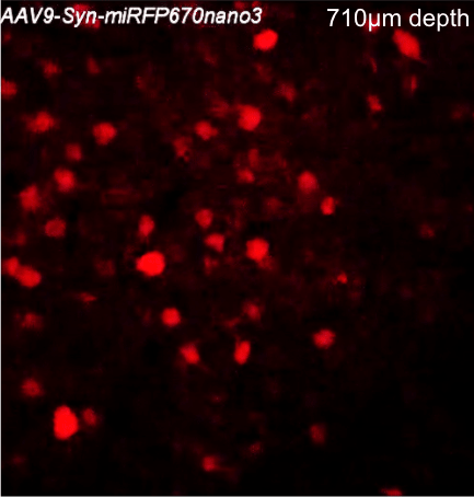
This post was contributed by guest blogger Daria Shcherbakova, a faculty member at Albert Einstein College of Medicine. Several sets of near-infrared fluorescent proteins (NIR FPs) and biosensors have been created recently. As developers of many of these probes, we decided to ...
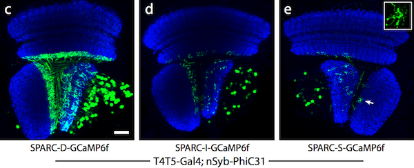
Until recently, there were no completely genetic tools that would allow researchers to label just a fraction of a single genetically-defined subset of cells. By labeling fewer cells in a population, it’s easier to visualize individual/non-overlapping cells. While transgenic ...
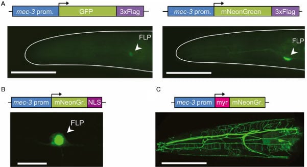
We are excited about our new partnership with Allele Biotechnology which allows researchers to deposit plasmids containing the fluorescent protein mNeonGreen. This fluorescent protein joins mTFP1 and mWasabi, as fluorophores from Allele Biotechnology that now can be deposited at ...
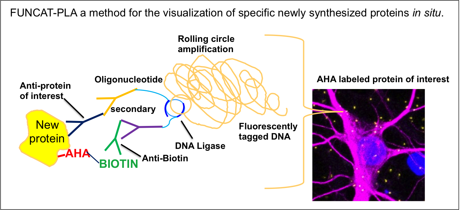
This post was contributed by guest blogger, Eugenia Rojas. A question worthy of a PhD: How do you visualize protein turnover within a neuron? For my PhD I studied a synaptic protein that is linked to neurodegeneration. The level of this protein is decreased in Alzheimer’s ...
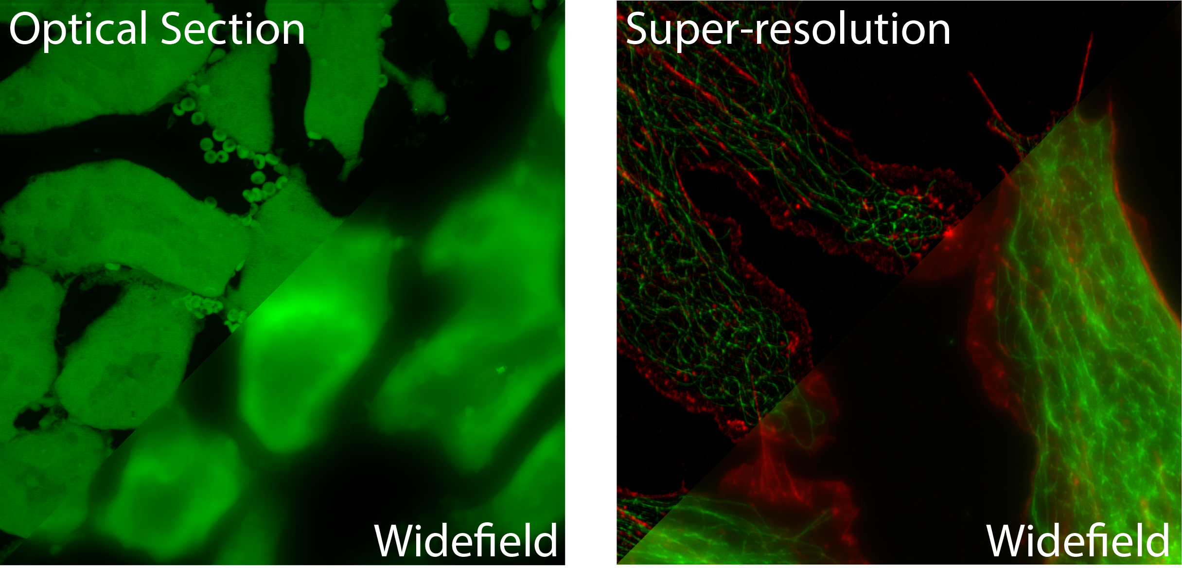
This post was contributed by Doug Richardson, Director of the Harvard Center for Biological Imaging and a Lecturer on Molecular and Cellular Biology at Harvard University. No matter whether you are a sports photographer at the Super Bowl, a medical technologist taking an x-ray, ...