Over 75,000 pathogenic genetic variants have been identified in humans and cataloged in the ClinVar database. Previously developed genome editing methods using nucleases and base editors have the potential to correct only a minority of those variants in most cell types. But ...
Deaminet 2026, held in Palm Springs in late January, brought together researchers studying deaminase enzymes across disciplines: from structural biologists resolving APOBEC proteins at atomic resolution, to cancer biologists dissecting mutational processes, to medical ...
It was huge news earlier this year: the first patient in the world, an infant, was successfully treated with a CRISPR gene editing therapy personalized to his genetic mutation. “Baby KJ” was diagnosed with a rare and dangerous metabolic disease shortly after his birth. Within a ...
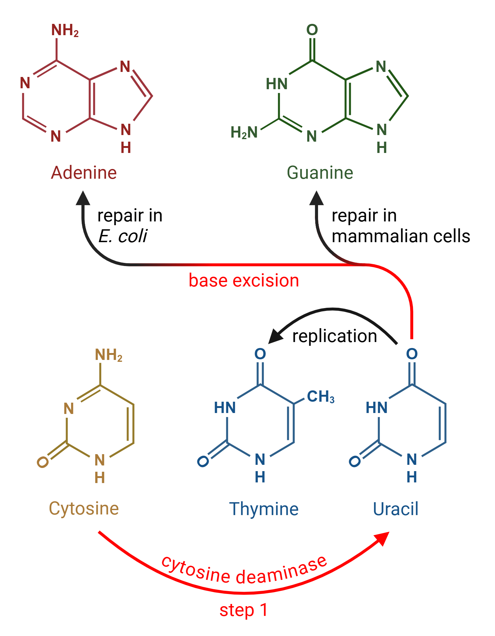
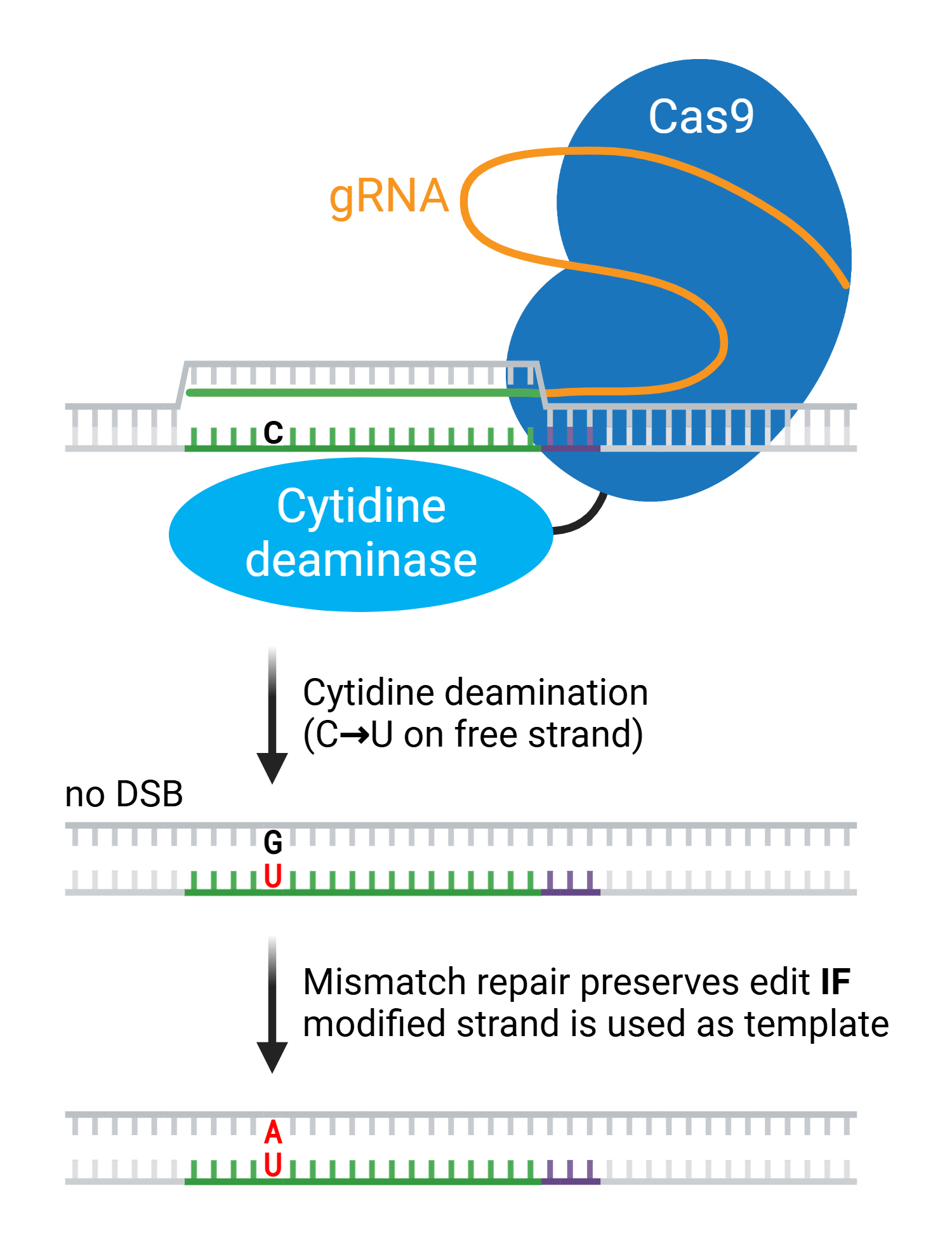
In our last post, we talked about the first base transversion editors: CGBEs, or C → G Base Editors. CGBEs first convert a cytosine (C) to uracil (U), just like Cytosine Base Editors (CBEs). But unlike CBEs, CGBEs then excise the U to create an abasic (empty) DNA site using ...
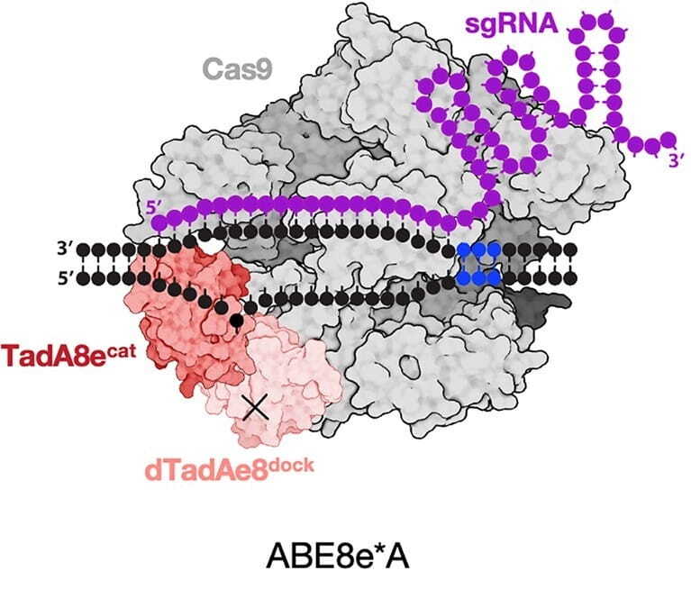
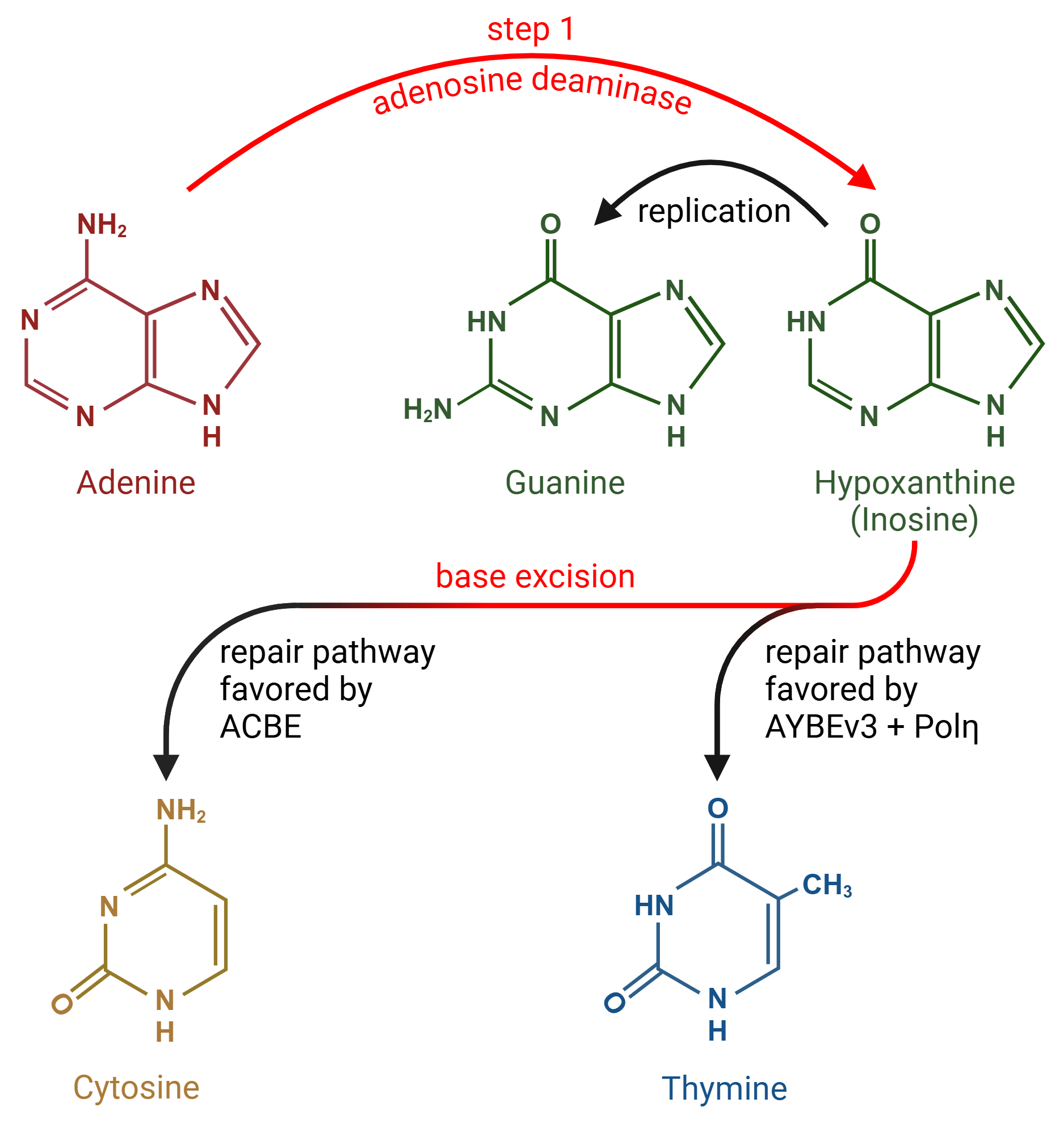
The first base editors revolutionized CRISPR gene editing. Cytosine base editors (CBEs) and adenine base editors (ABEs) chemically modify target bases without breaking the DNA backbone, making them efficient and precise tools for altering DNA sequences. These first base editors ...
Early CRISPR applications were often limited by the low editing efficiency of homology-directed repair (HDR), the pathway for resolving DNA double-strand breaks (DSBs) preferred by researchers. Compared to non-homologous end joining (NHEJ), HDR occurs at a relatively low ...
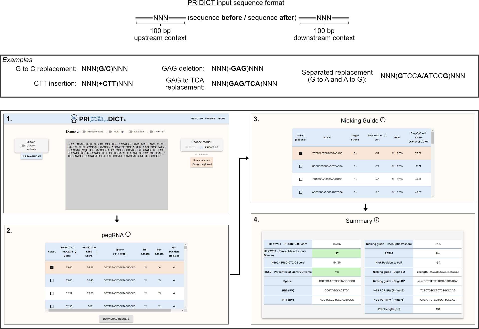
We recently updated our blog post on Prime Editing, and that meant rereading many of the original papers reporting various prime editing tools. These papers are chock full of great tips to guide your experimental design, especially the design of the RNA sequences you’ll use in ...
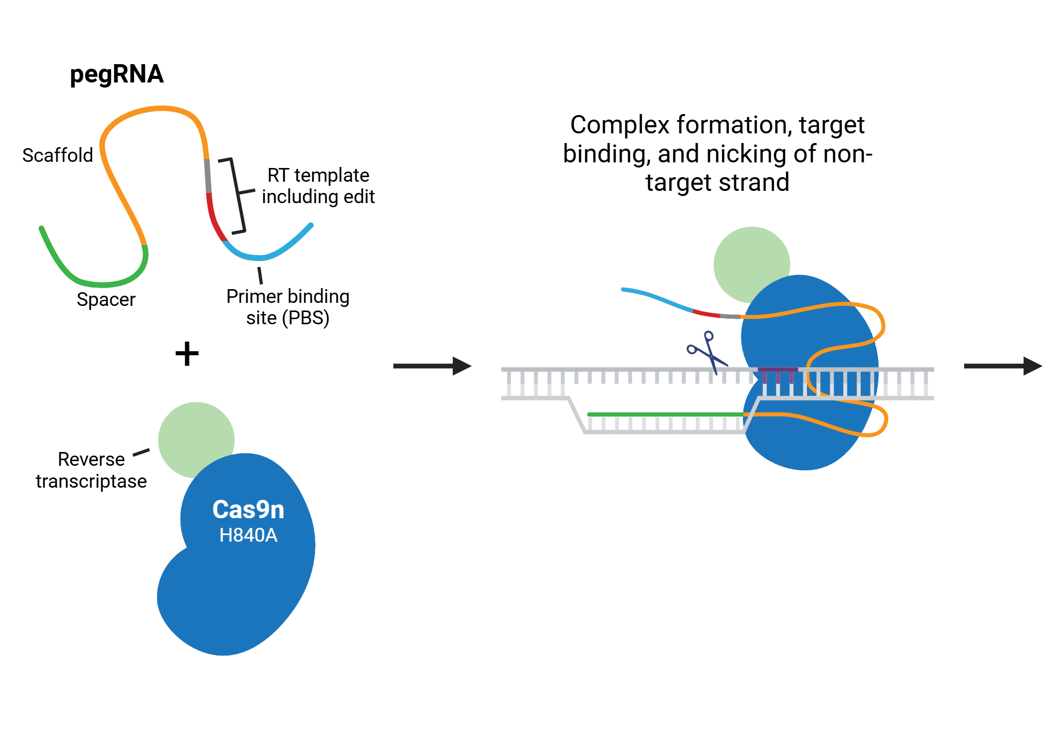
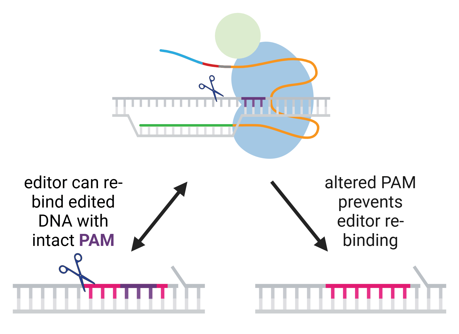
Prime editing is a versatile genome editing technology that allows precise modifications of DNA (replacements, small insertions, and deletions) without introducing DNA double-strand breaks (Anzalone et al., 2019; Chen & Liu, 2023). This method uses a prime editor (typically ...