Optogenetics gives you the power to control cells or organisms with the flip of a switch. You may be familiar with popular light-sensitive ion channels used to control activities like neuronal signaling — think of a mouse with an LED brain implant or a worm wiggling back and ...
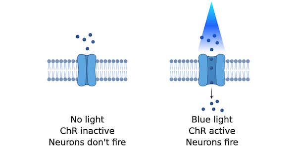
In 2005, Boyden et al. described the first light-activated tool for controlling neuronal activity, channelrhodopsin-2 (ChR2), a blue light-activated cation channel, from the archaebacteria Chlamydomonas reinhardtii. When exposed to blue light, this channel activates neuronal ...
🎶 “Bye bye L-arabinose drive Put your cultures in the shaker Turn the LEDs on And when you want You can just turn them off No need for any wash No need for any wash…” 🎶 - Barbara Di Ventura If you follow Barbara Di Ventura on Twitter, you might have seen the video of her ...
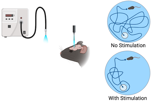
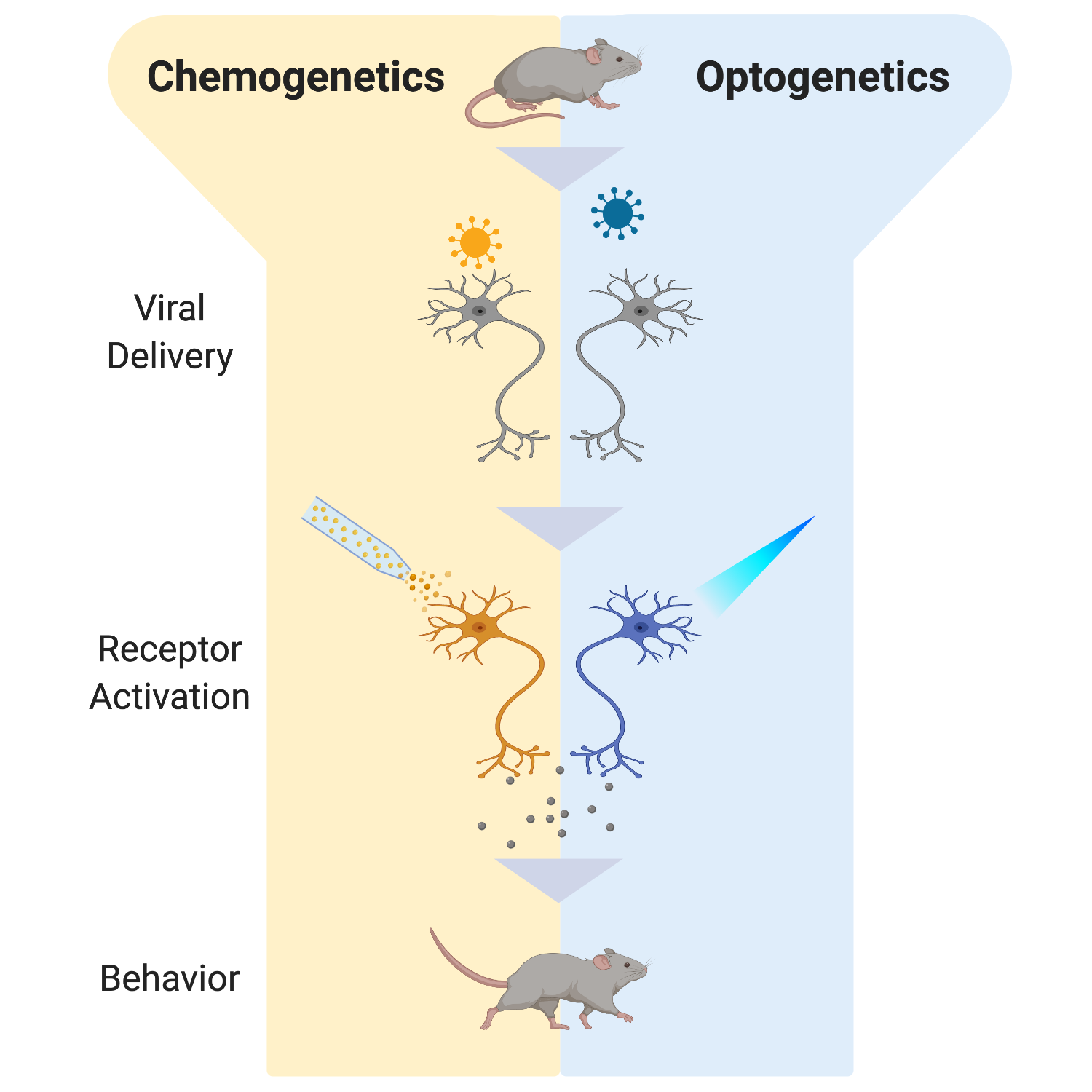
This post was contributed by Adriana Galvan, an associate professor at Emory University School of Medicine. Optogenetics and chemogenetics are powerful tools to modulate the activity of neurons and other brain cells. Since the opsins or chemogenetic receptors used in these ...
Optogenetics is a neuroscience method that lets you fire neurons with the flick of a light switch. Neurons are not typically persuaded to fire when light is shined on them, but the expression of light-gated ion channels such as channelrhodopsins (ChRs) makes them ...
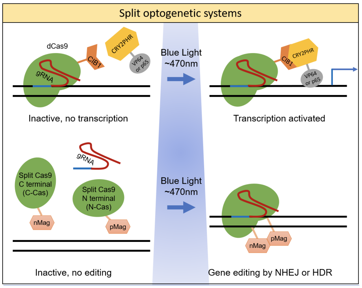
This blog post was originally written by Caroline LaManna, published Mar 8, 2016. The updated and expanded version by Nyla Naim was published Sept 3, 2020. Scientists around the world have been making major improvements to CRISPR technology since its initial applications for ...
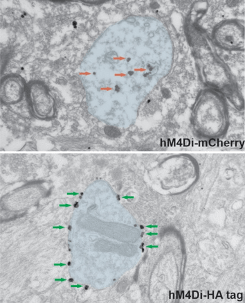
Chemogentic and optogenetic technologies have pushed the boundaries in neuroscience by granting targeted control over neuronal activity. While they serve similar purposes, both techniques offer researchers different advantages and limitations. The four main factors in which ...
It’s time to choose your own protein purification adventure. You want to purify your favorite protein (YFP). You have two options: Option #1: Affinity tag purification You tag YFP and use an affinity column for purification. After binding YFP to the column, you wash several ...