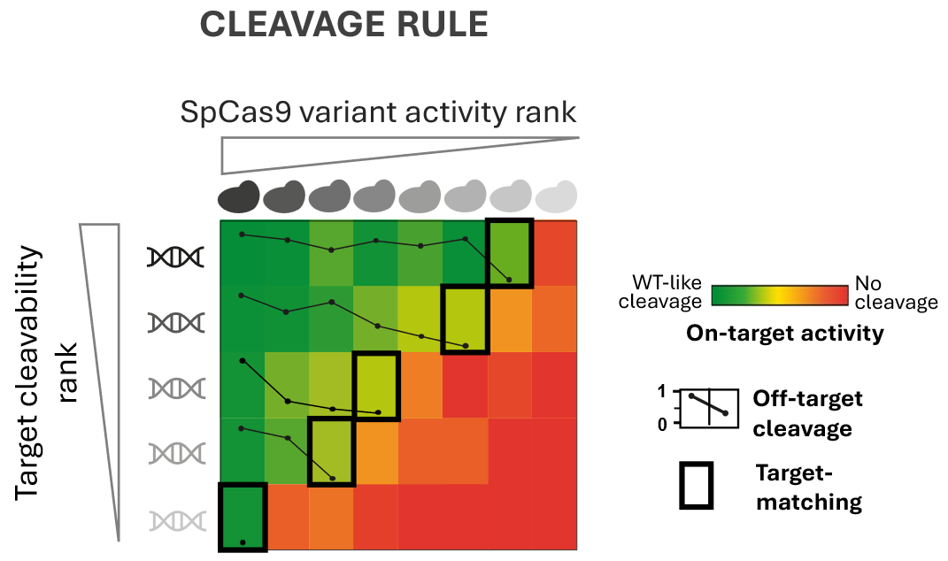
ByBalázs Csoma, Hungarian Research Network CRISPR nucleases are remarkably precise molecular tools for cutting DNA. But “remarkably precise” does not mean “perfectly specific.” In reality, CRISPR nucleases occasionally make mistakes and cleave DNA sequences that only resemble ...
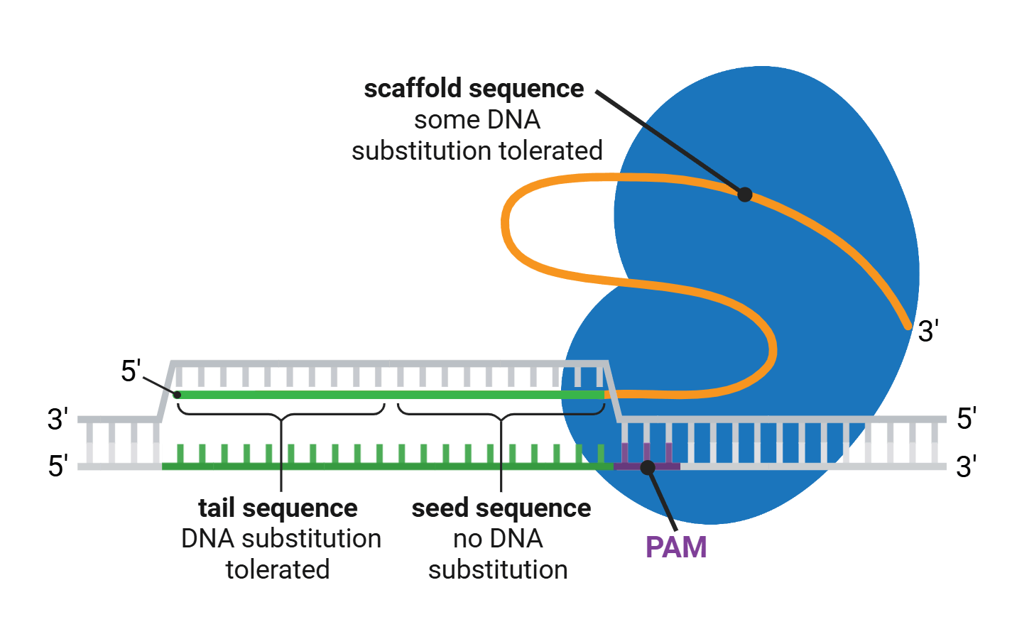
The genome-editing tool CRISPR is famously RNA-guided... except when it’s not. Turns out, carefully designed RNA-DNA hybrid strands work just as well—or maybe even better—at guiding Cas nucleases to specific genomic targets.
We recently updated our blog post on Prime Editing, and that meant rereading many of the original papers reporting various prime editing tools. These papers are chock full of great tips to guide your experimental design, especially the design of the RNA sequences you’ll use in ...
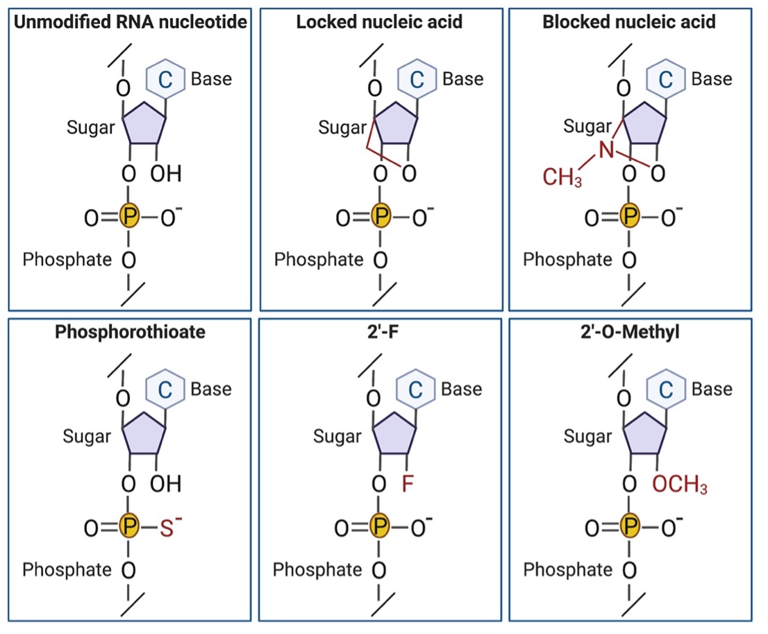
We’ve all heard “Get that tube on ice!!” and “I hope it isn’t degraded” when scientists talk about their precious RNA samples. RNA is inherently less stable than most macromolecules used in scientific research such as DNA or protein. It comes as no surprise then that stability ...
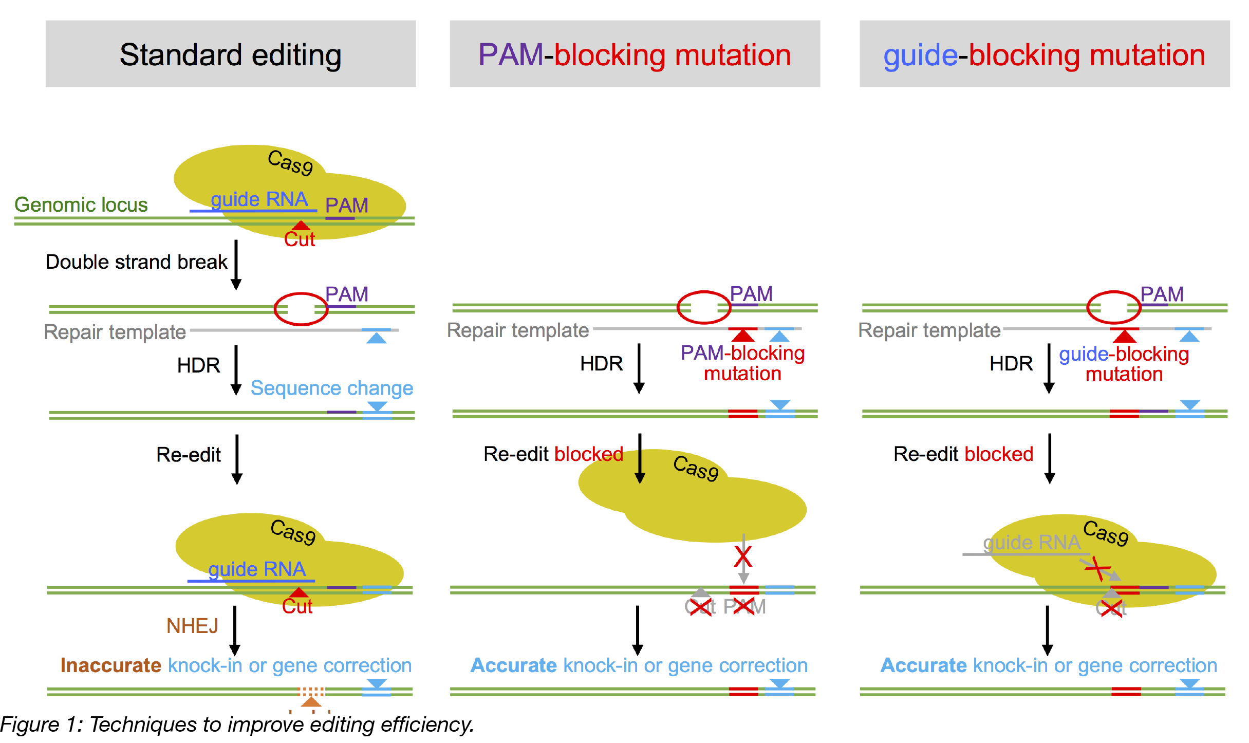
This post was contributed by guest bloggers Dominik Paquet and Dylan Kwart from Ludwig-Maximilians-University in Munich and Marc Tessier-Lavigne’s lab at the Rockefeller University in NYC. The CRISPR/Cas9 system is a versatile tool for precise gene editing in many organisms and ...
We all know that in the lab there are often little tricks that are essential for experiments but that nobody talks about. After months of troubleshooting, those people who did not tell you that essential thing ask incredulously, “You seriously didn’t add 3 microliters of 5 mM ...
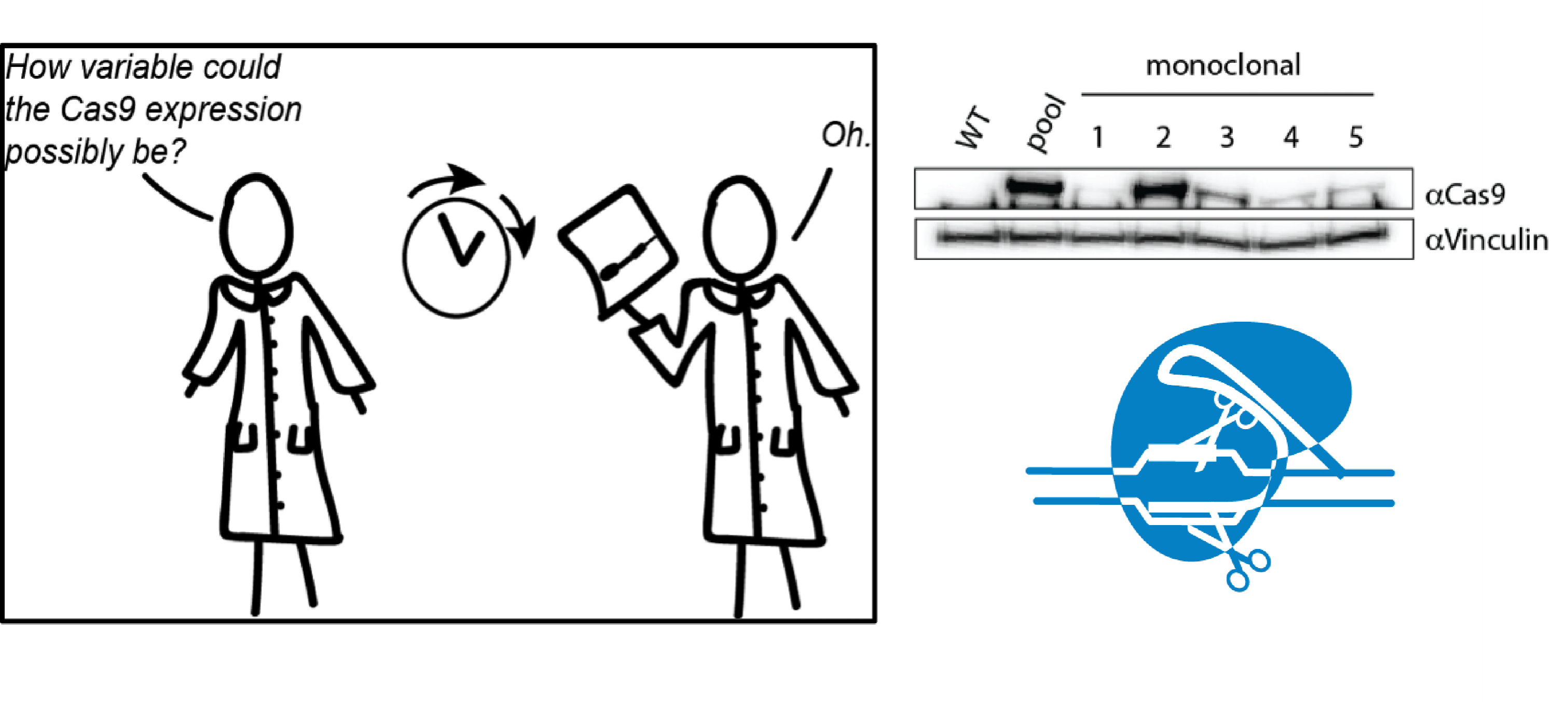
This post was contributed by guest blogger Søren Hough, a Biochemistry PhD candidate at the University of Cambridge. One of the most important steps in the CRISPR experimental process is validating edits. Regardless of which CRISPR genome editing system you use, there remains a ...
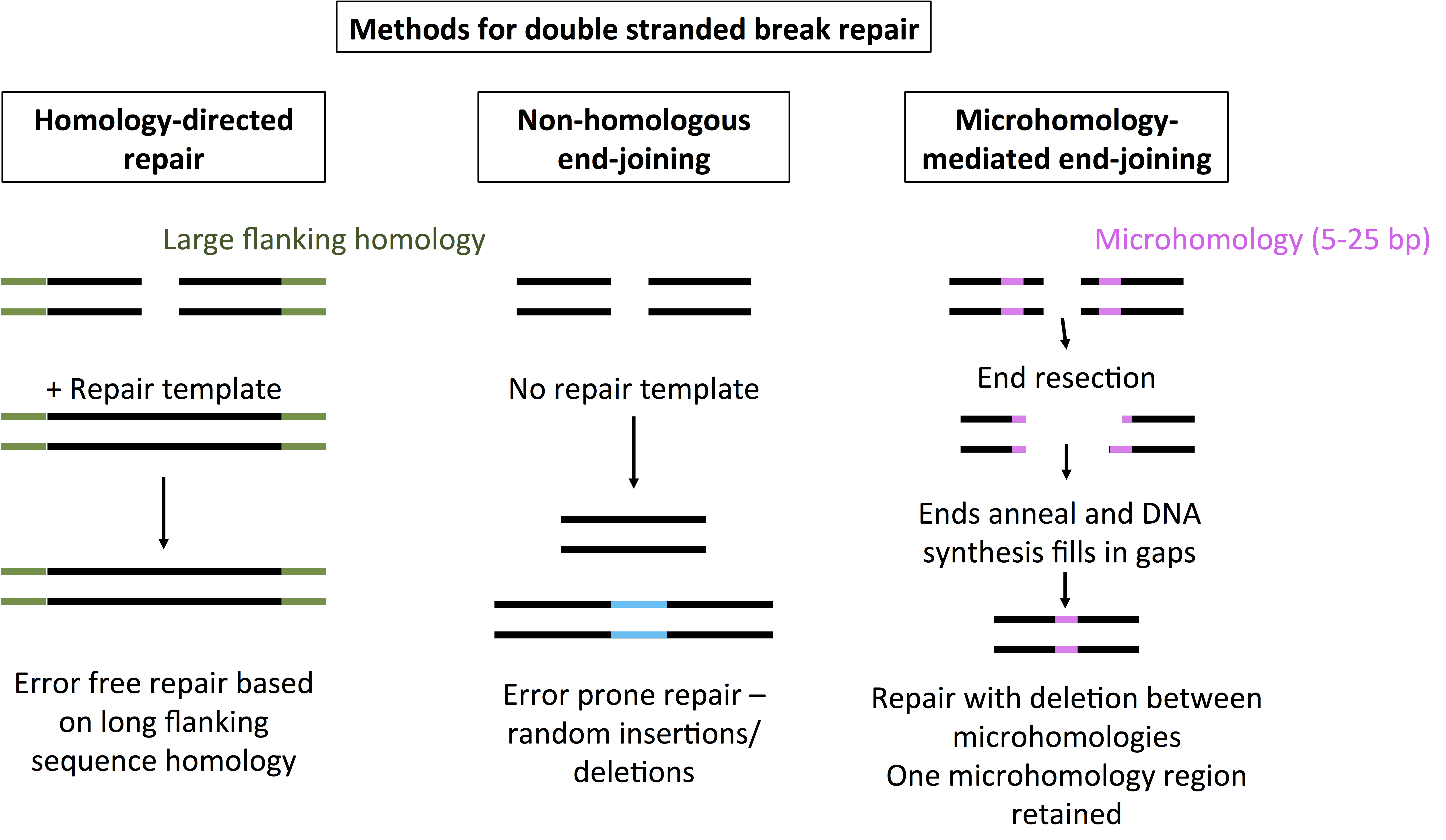
If you follow CRISPR research, you know all about using non-homologous end-joining (NHEJ) to make deletions or homology-directed repair (HDR) to create precise genome edits. But have you heard of another double-stranded break repair mechanism: MMEJ (microhomology-mediated ...