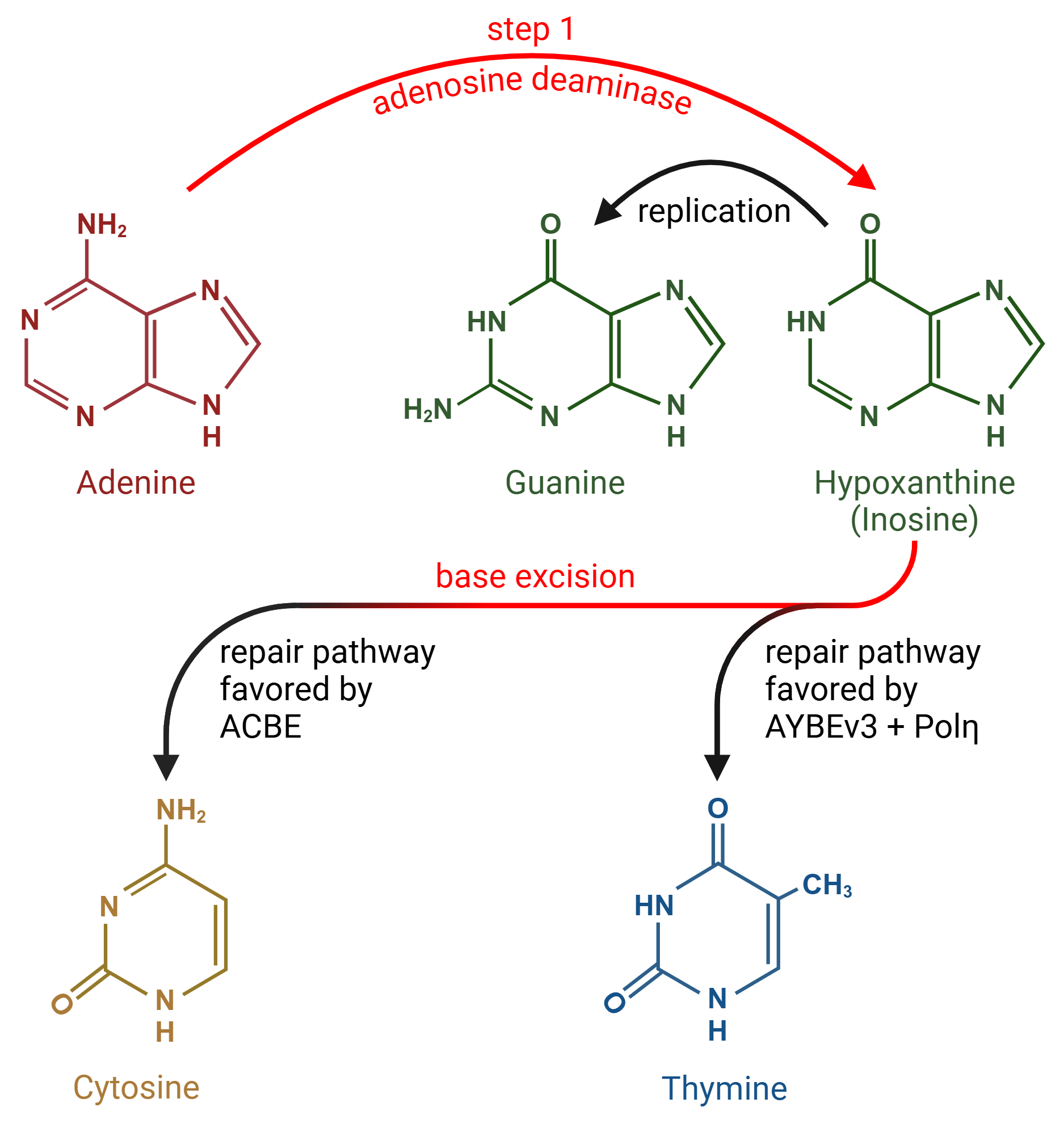
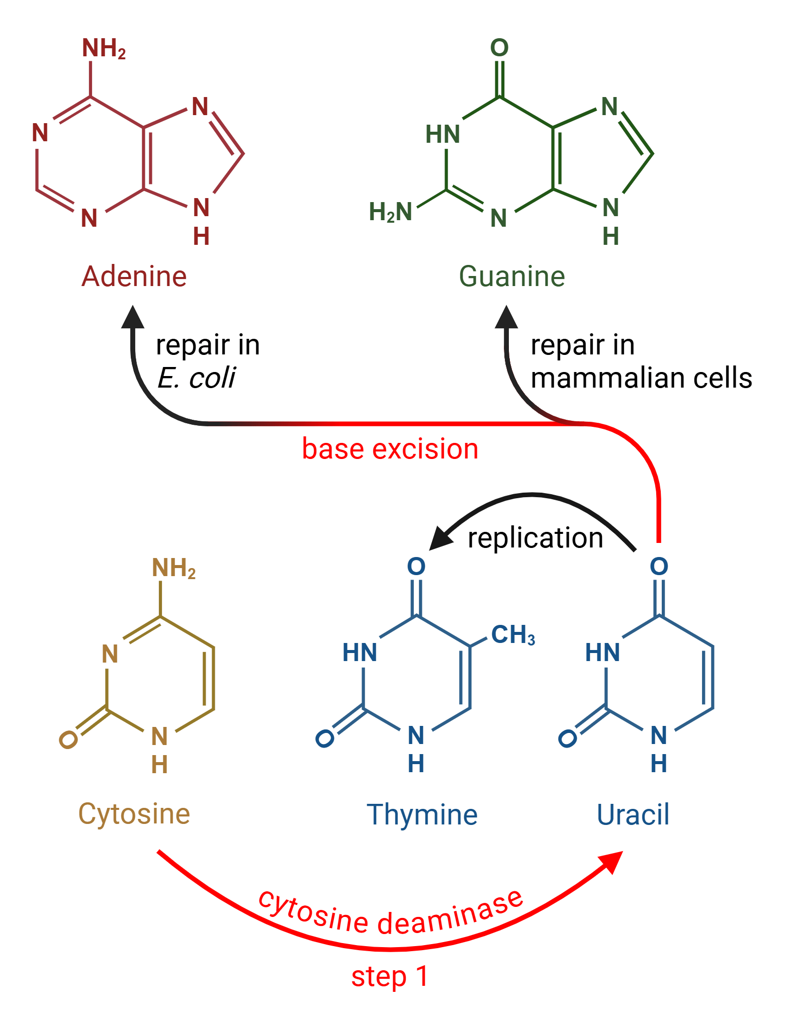
In our last post, we talked about the first base transversion editors: CGBEs, or C → G Base Editors. CGBEs first convert a cytosine (C) to uracil (U), just like Cytosine Base Editors (CBEs). But unlike CBEs, CGBEs then excise the U to create an abasic (empty) DNA site using ...
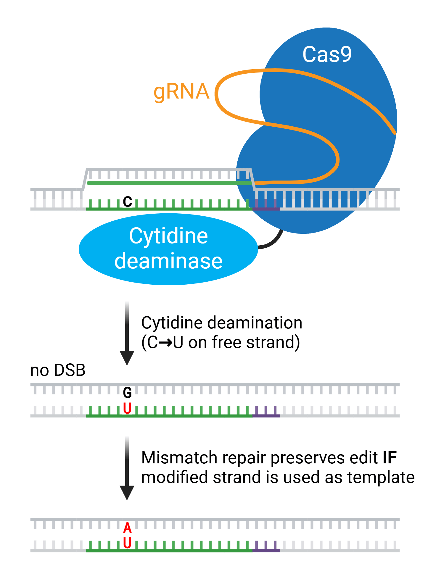
The first base editors revolutionized CRISPR gene editing. Cytosine base editors (CBEs) and adenine base editors (ABEs) chemically modify target bases without breaking the DNA backbone, making them efficient and precise tools for altering DNA sequences. These first base editors ...
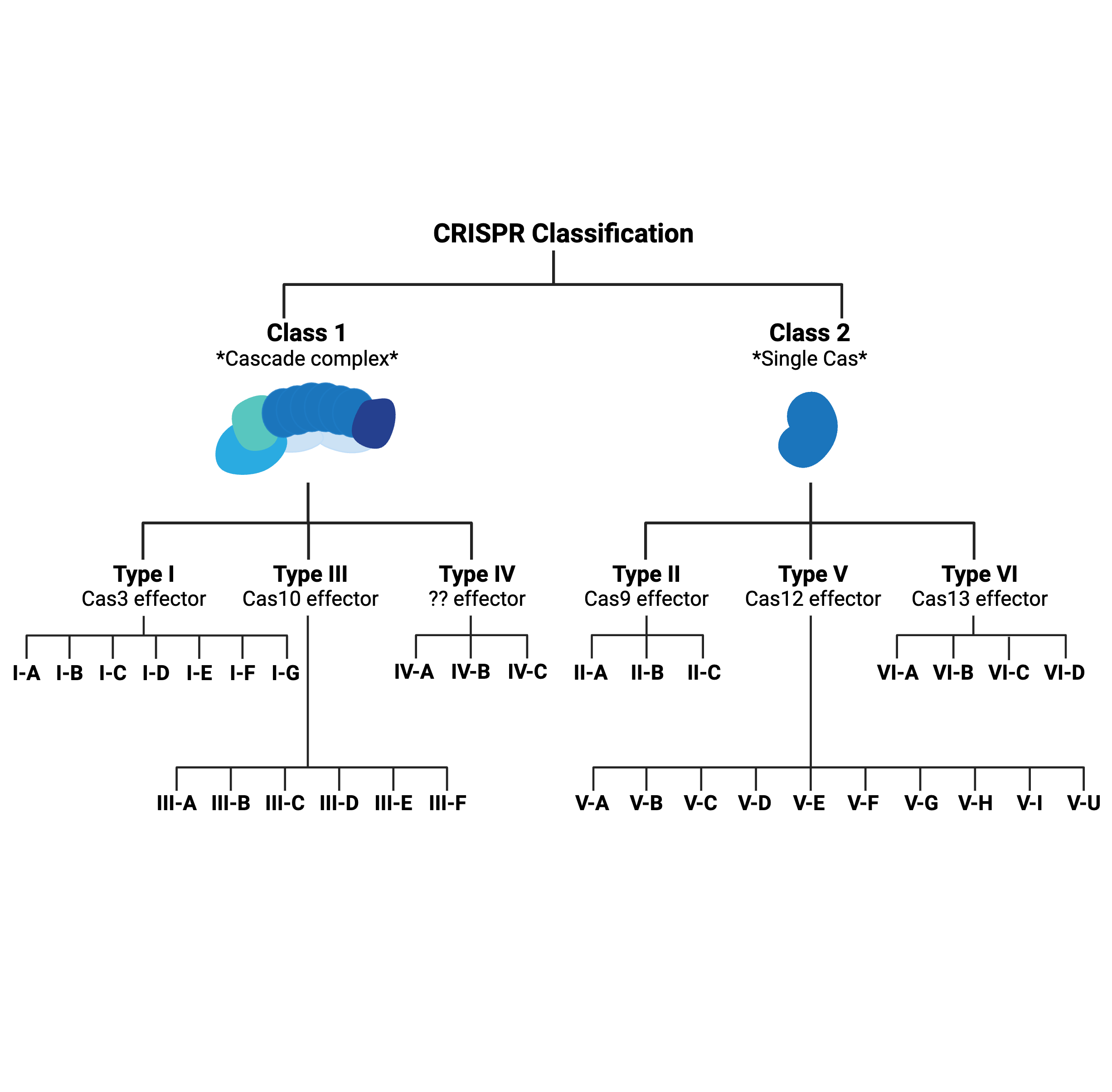
What’s in a type? That which we call CRISPR, by any other name would…probably still edit genomes.
Early CRISPR applications were often limited by the low editing efficiency of homology-directed repair (HDR), the pathway for resolving DNA double-strand breaks (DSBs) preferred by researchers. Compared to non-homologous end joining (NHEJ), HDR occurs at a relatively low ...
Over 75,000 pathogenic genetic variants have been identified in humans and cataloged in the ClinVar database. Previously developed genome editing methods using nucleases and base editors have the potential to correct only a minority of those variants in most cell types. But ...
You’ve probably heard that only 2% of our genome is made of protein-coding genes, and you might be wondering what the rest of our genome could possibly be made up of. The answer is… drum roll please… non-coding RNAs! You probably didn’t see that coming, right? Non-coding RNAs ...
CRISPR is a sleek acronym for a real mouthful of a phrase: Clustered Regularly Interspaced Short Palindromic Repeats. That contrast of simplicity and complexity is reflected in the biology, too. CRISPR is an elegant bacterial immune system and an efficient gene editing tool… but ...
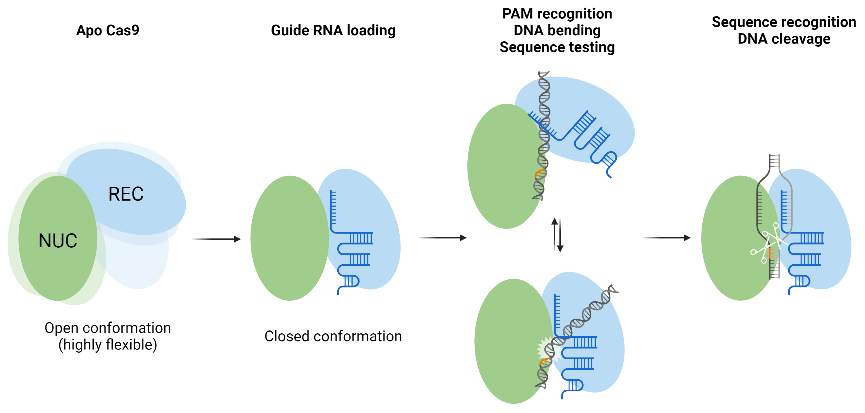
Have you ever designed a CRISPR guide RNA and wondered why it is limited to only 20 bases, or why it’s so important to choose a target sequence with a nearby protospacer-adjacent motif (PAM)? Cas9 is becoming an ever more ubiquitous tool for genome engineering, and studying its ...


-min.png)