The calcium is strong here….I can sense it….sound like a plot to an biology scifi movie? Or maybe it’s a biosensor?! You can do more than just ‘sense’ biological compounds and reactions; you can quantify them with biosensors! In this blog we will review the basics of biosensors and how to use viruses to set up these systems.
Components of a biosensor
Some of the biosensors used in research are very sophisticated, but at their core they can all be boiled down pretty simply – they are systems that measure biological phenomena by generating an output which is directly proportionate to the amount of a given factor within a biological system. Basically, biosensors provide quantifiable readouts for levels of different chemicals, hormones, proteins, etc., that you are interested in.
The most obvious component of a biosensor is the thing that you care about within your cellular or animal system. This ‘thing’ could be a neurotransmitter, such as norepinephrine, or an electrolyte such as calcium. This varied component, which we will refer to as the analyte, needs to be specifically recognized by another factor, the receptor. The receptor could be something like an antibody, enzyme, DNA, etc. The receptors job is to generate a quantitative signal upon recognizing the analyte. This signal depends on the system and the receptor, but readouts could involve fluorescence, pH change, heat, etc. The last component of the biosensor is the signal transducer which will quantitatively give a readout for whatever the bio-recognition event is (fluorescence, pH, etc.). The transducer will generate a numerical value which represents the amount of analyte present in the system at a given point in time. For example, if ATP was the molecule of interest (analyte), and accumulation of ATP activated GFP expression (receptor), then the transducer could be a microscope, plate reader, or any other instrument which measured GFP levels and converted that signal into a readable value.
Types of biosensors
So now that we know what makes up a biosensor, we can discuss what flavors of sensors exist and are ready-to-use. While biosensors span the gamut of applications within chemistry, biology, and physics, in this blog we will focus on those which can be applied in cell culture and animal models primarily.
Fun fact: Almost all of the biosensors discussed are genetically encoded, and luckily, we have pre-designed plasmids for these systems!
Ion sensors
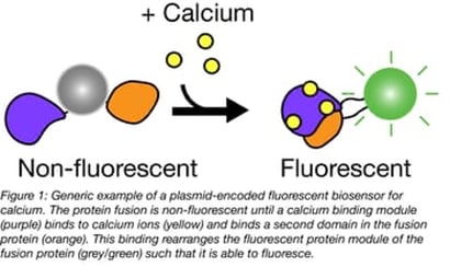
One common biosensor class is designed to monitor metal ions. Within these ions, calcium is by far one of the most popular, with dozens of sensors developed for it. Calcium triggers muscle contraction, and calcium channels are activated for neuronal action potentials; it’s an ion of interest for many disciplines! Most of the sensors designed for calcium generate a fluorescent output, which will turn on, change color, or change intensity of fluorescence in the presence of calcium. This includes FRET-based sensors such as Twitch. While calcium is king in the sensor world, there are sensors available for other ions including magnesium, potassium, and zinc.
These ion sensors can be targeted to different cellular locations or tissues depending on the biology you are studying. For example, calcium dynamics in the cytosol and sarcoplasmic reticulum are of interest to muscle biologists, while neuroscientists would like to monitor calcium specifically in their favorite neurons. This specificity is feasible partially because of viruses—keep reading to find out how!
 |
Neurotransmitters
Many canonical fluorescent as well as FRET-based biosensors have been engineered to detect a wide array of neurotransmitters ranging from dopamine to glutamate, all the way to acetylcholine. These sensors have been tested in vivo, in tissue preps, in multiple species, and in cell culture models. Some of them have even been expanded for multiplexed imaging for advanced monitoring of different factors, in different or the same cellular location, all at once!
Small molecules
What type of small molecules can you monitor with biosensors? You can keep tabs on my personal favorite (the caffeine of the cell!)—ATP. There are biosensors for intracellular, extracellular, and even the ADP:ATP ratio. Most of these sensors are fluorescence-based and have been optimized for a wide-range of uses to monitor the fundamental cellular building blocks.
Physiological factors
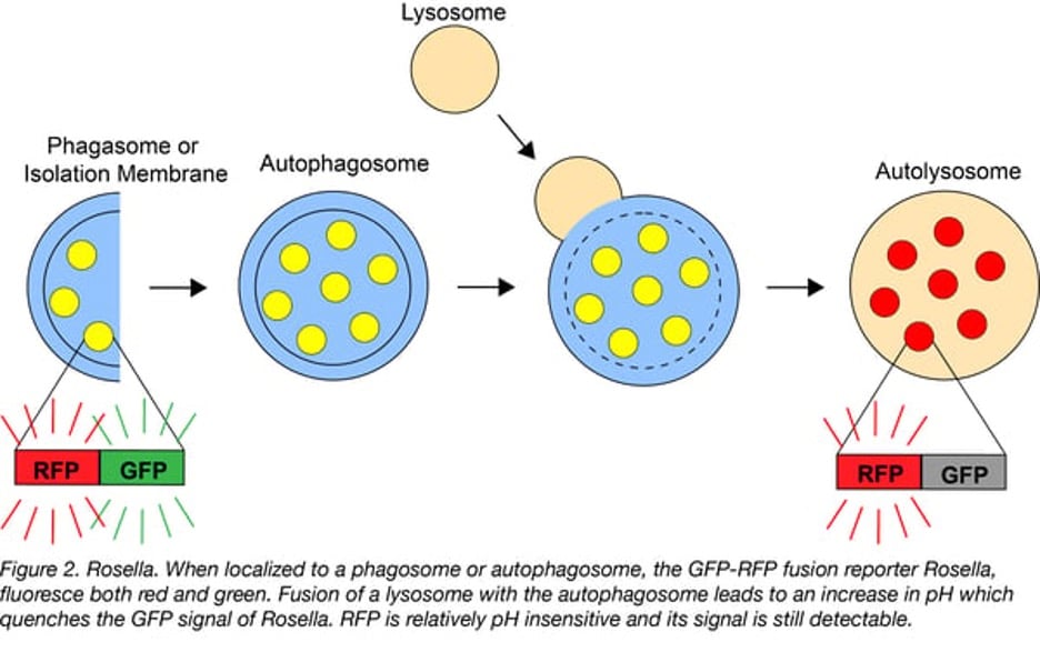
Unlike the previously described sensors, this class measures either factors as proxies for a cellular process or directly measure a cellular process. Cellular phenomena such as exocytosis, autophagy, voltage, and tension all have specific sensors designed for them.
For example, this class includes tension monitors associated with specific proteins of interest such as actinin. When tension is high vs. low in the cell the donor and acceptor of a FRET pair on actinin will produce different signals based on changes to the structure of actinin in low vs. high tension. These systems are powerful tools to measure real-time cellular reactions to stimuli and as well as changes in cellular dynamics during so-called housekeeping activities.
 |
Viruses and biosensors
So how do viruses factor into all of these diverse biosensors? Well, a lot of these sensors are already engineered into viral vectors, so if you want to use them, you’ll need to use the vectors (or redesign the tools). It’s a good thing, though! Viral delivery has several advantages over other mechanisms. For one, many viral platforms, such as AAV or retrovirus, can either permanently integrate and/or express long term (up to several years!). Biosensors tend to be more reproducible when used as stable systems, rather than transiently overexpressing at the time of use.
Another great viral perk for delivery is targetability. As mentioned prior, many biosensors need to be in specific tissues or cells to monitor their target analyte. This can be difficult to achieve in whole organisms, but AAVs make it possible. In AAV systems, different serotypes can be used to affect or determine viral tropism—the type of cells the viral particles infect. This can direct your biosensor to specific tissues or cell (sub)types for expression and downstream monitoring.
Nervous about making a virus for your biosensor? Don’t be! We have you covered with viral resources and safety tips. Still worried? We make virus with some of our most popular biosensors already packaged and ready to deliver.
Ready to give biosensors a try? Peruse our available biosensors or build your own! When you’re ready to deliver your sensor, whether you choose lentivirus, gamma retrovirus, or AAV as your vehicle, we’ve got you covered with packaging plasmids!
References and Resources
References
Bhalla N., Jolly P., et al. Introduction to Biosensors. Essays Biochem. 2016 Jun 30;60(1):1-8. DOI: 10.1042/EBC20150001
Leopold, A. V., Shcherbakova D. M., Verkhusha, V. V., Fluorescent Biosensors for Neurotransmission and Neuromodulation: Engineering and Applications. Frontiers in Cellular Neuroscience. 2019 Oct Vol. 13; 1662-5102. DOI: 10.3389/fncel.2019.00474
Resources on Addgene.org
Resources on the Addgene blog
Topics: Viral Vectors


Leave a Comment