Using AAV vectors in complex biological systems can be tricky at best, and downright infuriating at worst. While it is tempting to just dive right in and start injecting your virus, a successful AAV experiment starts with validation and optimization. Although there are different ways to ultimately achieve success, we find it is usually faster and easier to do the validation first.
AAV-mediated gene expression can differ extensively depending on the experimental system used, so you’ll need to know what expression pattern you need, and the factors in your system that can affect expression, before starting the selection process. Factors to think about are (1) your experimental conditions and measurements; (2) the cells you’re targeting; (3) how much expression is needed to read your experimental output; and (4) the consequences to your experiment if there’s off-target expression.
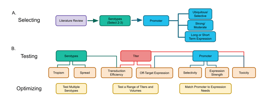
Once you know that, you’re ready to start selecting, testing, and optimizing three key variables of an AAV vector: serotype, promoter, and titer.
|
| Figure 1: Flow chart depicting a) steps for selecting serotypes and promoters to test in your AAV system and b) steps for testing and optimizing your serotypes, titer, and promoter. Note that several steps in testing are effected by multiple AAV variables. |

Serotypes
Serotype refers to the different protein compositions of AAV capsids, which determine how a virus enters the cell. Different serotypes utilize a number of different receptors, co-receptors, and other mechanisms to gain entry into the cell (Dudek et al., 2018; Korneyenkov & Zamyatnin, 2021), meaning what works great in one system or cell type can completely fail in a different system (Issa et al., 2023). This can be particularly burdensome in the brain where many different cell types, or even different phenotypes of the same cell type, are intermingled in the same brain nucleus: a serotype that works great in a GABA neuron, for instance, may not transduce the adjacent norepinephrine neuron at all. There may be more than one serotype that will efficiently transduce your cells. In fact, we usually recommend testing more than one.
Most AAV serotypes have broad tropism (the ability of a virus to enter a particular cell type), but there are differences in tropism between animal species or even strains of the same species. What works well in a mouse may not work as well in a rat or non-human primate. Likewise, in vitro efficacy doesn’t always translate to in vivo efficacy. You’ll therefore need to test potential AAV vectors in your specific experimental system, rather than relying on other systems as proxies.
Finally, the size and receptor interactions of a serotype can affect spread from the injection site (Kanaan et al., 2017), axonal transport (Murlidharan et al., 2014), and postsynaptic transmission (Oh et al., 2020). For example, AAV1 and AAV9 tend to spread farther from the injection site than AAV2 (Watakabe et al., 2015) (Figure 1). You’ll need to consider if such properties could help, or harm, your experimental goals when designing and testing your AAV.
-min.png?width=569&height=461&name=AAV101_Preparing_Figure%201_2%20(1)-min.png) |
| Figure 2: Comparison of AAV serotypes 1, 2, 5, 6, 8, and 9 in two different brain regions. Image modified from Aschauer et al., 2013, Figure 1. Used under Creative Commons Attribution License. |
Within the nervous system, remember that some serotypes can be transported retrogradely and/or anterogradely along the axon. Others, like AAV1 and AAV9, can also transduce postsynaptic cells when used at very high titers (Zingg et al., 2017), which can lead to cells in far away brain regions expressing your gene of interest. This can cause issues in tracing studies, where it could potentially label cells not directly connected to the injection site, or when using chemogenetic tools, where modification of cellular activity outside of your experiment site or circuit can occur.
Promoters
The promoter controls how the gene of interest will be expressed; it can be ubiquitous, cell-type selective, or inducible.
Ubiquitous promoters
These often-strong promoters offer the most flexibility for expression in a large number of cell types and are useful if you're going to be working in several different systems or want to transduce a large number of different cells. Promoters such as chicken-beta-actin or CAG can drive strong early gene expression, but can become methylated over time, resulting in decreased expression (Damdindorj et al., 2014; Nieuwenhuis et al., 2021). It is important to note that this expression pattern can be affected by the cell and/or tissue type, so you’ll need to test for this in your system if you’re measuring expression over a period of weeks (in vitro) or months (in vivo).
Selective promoters
Selective promoters, such as synapsin, glial fibrillary associated protein (GFAP), or tyrosine-hydroxylase (TH), can limit which cells express your gene. Off-target expression can still occur (Fig. 3) - for example, some neurons can transiently express TH - so you’ll need to check for this in your system and then test each vector before starting your experiments (Rolland et al., 2016; Su et al., 2004; Taschenberger et al., 2017).
-min.png?width=563&height=524&name=AAV101_Preparing_Figure%202%20(1)-min.png)
|
| Figure 3: Comparison of two astrocyte selective promoters demonstrating off-target expression in both oligodendrocytes (Olig2) and neurons (NeuN). Image from Griffin et al., 2019, Figure 4. Used under Creative Commons Attribution 4.0 International License. |
Inducible promoters
This type of promoter allows you to decide when to turn on, or turn off, gene expression by administering an inducing agent like doxycycline (Favre et al., 2002). They are useful when you need to control the timing of expression in your experiment. Some inducible constructs can exhibit leaky expression in the absence of the inducing drug, so be sure to test for this when validating your viral vector.

Titer
Once you have selected potential serotypes and promoters to begin testing, you’ll next need to decide how much virus to put into your system, which is controlled through titer (concentration, given as viral genome copies/mL (GC/mL)), and total injection volume.
You’ll often hear that “high titer” is required for efficient AAV transduction, usually without any numbers for context. That’s because the number can vary depending on your route of administration, what cells you want to deliver to, and how you’ll be measuring your experimental output. You’ll also want to keep in mind the serotype you’ve picked, since that can affect the spread of the virus.
A better way to define high titer, then, is this: It’s the optimal number of viral particles per volume required to express the gene of interest, in the cells being studied, at an adequate level for your experimental measurements. The number itself is determined through validation and optimization of your expression system for both the titer and total injection volume.

There are a few factors to consider. First, the titer required is inversely related to transduction efficacy: the higher the efficiency, the lower the titer and vice versa. The total volume needed, however, increases with the size of the region you want to cover - and as your titer changes, so may your needed volume (de Backer et al., 2010).
Second, high efficiency transduction coupled with a strong promoter, may result in overexpression of your gene and could lead to toxicity or trouble interpreting your results, while a low transduction efficiency and a weak promoter may result in unacceptably low expression.
Finally, the risks of potential toxicity and/or off-target effects (e.g. recombinase-dependent vectors) increases with the amount of virus you add, so it is strongly recommended to optimize to the “lowest number required” instead of using the first amount that looks effective. Ultimately, the perfect “high titer” is one that optimizes gene expression where you want it and minimizes expression where you don’t.
The key to good AAV selection
…is truly validation and optimization! Understanding serotypes, promoters, and titers will allow you to efficiently move through this process. Many other factors can also affect your AAV experiments. Luckily, the other posts in our Viral Vectors 101 series can help guide you with these, so keep reading!

Good luck, and happy optimizing!
Recommended Reading
Issa, S. S., Shaimardanova, A. A., Solovyeva, V. V., & Rizvanov, A. A. (2023). Various AAV Serotypes and Their Applications in Gene Therapy: An Overview. Cells, 12(5), 1–41. https://doi.org/10.3390/cells12050785
Murlidharan, G., Samulski, R. J., & Asokan, A. (2014). Biology of adeno-associated viral vectors in the central nervous system. Frontiers in Molecular Neuroscience, 7. https://doi.org/10.3389/fnmol.2014.00076
References and Resources
References
Aschauer, D. F., Kreuz, S., & Rumpel, S. (2013). Analysis of Transduction Efficiency, Tropism and Axonal Transport of AAV Serotypes 1, 2, 5, 6, 8 and 9 in the Mouse Brain. PLOS ONE, 8(9), e76310. https://doi.org/10.1371/journal.pone.0076310
Damdindorj, L., Karnan, S., Ota, A., Hossain, E., Konishi, Y., Hosokawa, Y., & Konishi, H. (2014). A comparative analysis of constitutive promoters located in adeno-associated viral vectors. PLoS ONE, 9(8), 1–10. https://doi.org/10.1371/journal.pone.0106472
de Backer, M. W. A., Brans, M. A. D., Luijendijk, M. C., Garner, K. M., & Adan, R. A. H. (2010). Optimization of Adeno-Associated Viral Vector-Mediated Gene Delivery to the Hypothalamus. Human Gene Therapy, 21(6), 673–682. https://doi.org/10.1089/hum.2009.169
Dudek, A. M., Pillay, S., Puschnik, A. S., Nagamine, C. M., Cheng, F., Qiu, J., Carette, J. E., & Vandenberghe, L. H. (2018). An Alternate Route for Adeno-associated Virus (AAV) Entry Independent of AAV Receptor. Journal of Virology, 92(7). https://doi.org/10.1128/jvi.02213-17
Favre, D., Blouin, V., Provost, N., Spisek, R., Porrot, F., Bohl, D., Marmé, F., Chérel, Y., Salvetti, A., Hurtrel, B., Heard, J.-M., Rivière, Y., & Moullier, P. (2002). Lack of an Immune Response against the Tetracycline-Dependent Transactivator Correlates with Long-Term Doxycycline-Regulated Transgene Expression in Nonhuman Primates after Intramuscular Injection of Recombinant Adeno-Associated Virus. Journal of Virology, 76(22), 11605–11611. https://doi.org/10.1128/JVI.76.22.11605-11611.2002
Griffin, J. M., Fackelmeier, B., Fong, D. M., Mouravlev, A., Young, D., & O’Carroll, S. J. (2019). Astrocyte-selective AAV gene therapy through the endogenous GFAP promoter results in robust transduction in the rat spinal cord following injury. Gene Therapy, 26(5), Article 5. https://doi.org/10.1038/s41434-019-0075-6
Issa, S. S., Shaimardanova, A. A., Solovyeva, V. V., & Rizvanov, A. A. (2023). Various AAV Serotypes and Their Applications in Gene Therapy: An Overview. Cells, 12(5), 1–41. https://doi.org/10.3390/cells12050785
Kanaan, N. M., Sellnow, R. C., Boye, S. L., Coberly, B., Bennett, A., Agbandje-McKenna, M., Sortwell, C. E., Hauswirth, W. W., Boye, S. E., & Manfredsson, F. P. (2017). Rationally Engineered AAV Capsids Improve Transduction and Volumetric Spread in the CNS. Molecular Therapy - Nucleic Acids, 8, 184–197. https://doi.org/10.1016/j.omtn.2017.06.011
Korneyenkov, M. A., & Zamyatnin, A. A. (2021). Next Step in Gene Delivery: Modern Approaches and Further Perspectives of AAV Tropism Modification. Pharmaceutics, 13(5), 750. https://doi.org/10.3390/pharmaceutics13050750
Murlidharan, G., Samulski, R. J., & Asokan, A. (2014). Biology of adeno-associated viral vectors in the central nervous system. Frontiers in Molecular Neuroscience, 7. https://doi.org/10.3389/fnmol.2014.00076
Nieuwenhuis, B., Haenzi, B., Hilton, S., Carnicer-Lombarte, A., Hobo, B., Verhaagen, J., & Fawcett, J. W. (2021). Optimization of adeno-associated viral vector-mediated transduction of the corticospinal tract: Comparison of four promoters. Gene Therapy, 28(1–2), 56–74. https://doi.org/10.1038/s41434-020-0169-1
Oh, J. Y., Han, J. H., Lee, H., Han, Y. E., Rah, J. C., & Park, H. (2020). Labeling dual presynaptic inputs using cFork anterograde tracing system. Experimental Neurobiology, 29(3), 219–229. https://doi.org/10.5607/en20006
Rolland, A.-S., Kareva, T., Yarygina, O., Kholodilov, N., & Burke, R. E. (2016). Expression mediated by three partial sequences of the human tyrosine hydroxylase promoter in vivo. Molecular Therapy - Methods & Clinical Development, 3, 16062. https://doi.org/10.1038/mtm.2016.62
Su, M., Hu, H., Lee, Y., D’Azzo, A., Messing, A., & Brenner, M. (2004). Expression specificity of GFAP transgenes. Neurochemical Research, 29(11 SPEC. ISS.), 2075–2093. https://doi.org/10.1007/s11064-004-6881-1
Taschenberger, G., Tereshchenko, J., & Kügler, S. (2017). A MicroRNA124 Target Sequence Restores Astrocyte Specificity of gfaABC1D-Driven Transgene Expression in AAV-Mediated Gene Transfer. Molecular Therapy - Nucleic Acids, 8, 13–25. https://doi.org/10.1016/j.omtn.2017.03.009
Watakabe, A., Ohtsuka, M., Kinoshita, M., Takaji, M., Isa, K., Mizukami, H., Ozawa, K., Isa, T., & Yamamori, T. (2015). Comparative analyses of adeno-associated viral vector serotypes 1, 2, 5, 8 and 9 in marmoset, mouse and macaque cerebral cortex. Neuroscience Research, 93, 144–157. https://doi.org/10.1016/j.neures.2014.09.002
Zingg, B., Chou, X. lin, Zhang, Z. gang, Mesik, L., Liang, F., Tao, H. W., & Zhang, L. I. (2017). AAV-Mediated Anterograde Transsynaptic Tagging: Mapping Corticocollicular Input-Defined Neural Pathways for Defense Behaviors. Neuron, 93(1), 33–47. https://doi.org/10.1016/j.neuron.2016.11.045
More resources on the Addgene blog
Viral Vectors 101: An Introduction to AAV
Viral Vectors 101: The AAV Retrograde Serotype
Viral Vectors 101: Viral Applications
Topics: Viral Vectors, Viral Vectors 101, AAV


Leave a Comment