Antibodies are a go-to tool for detecting a protein of interest in cells and tissues. Although antibody production is well established, it’s also a process that’s difficult for individual labs to complete. The nanobody based RANbody platform from the Sanes Lab overcomes this limitation and allows for the flexible design and small scale production of antibodies.
Problems with antibodies
- Conventional antibodies are a non-renewable reagent - Their production requires animal sacrifice and because they are an animal derived product, there can be lot-to-lot variability in activity.
- No Genetic Tagging - Since antibodies are produced in animals instead of via recombinant DNA, they can’t be genetically tagged and instead are conjugated to reporters (i.e. fluorescent dyes, HRP, etc. ) after purification.
- Antibodies are difficult for an individual lab to produce - Hybridomas offer an alternative and renewable source of monoclonal antibodies, but they are laborious to generate and costly to share.
What is a RANbody?
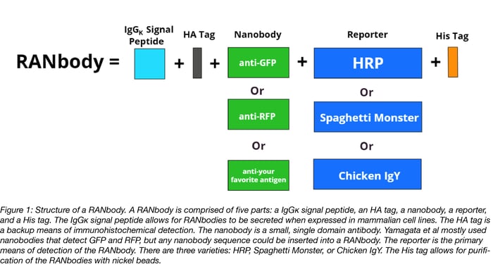
A RANbody is a fusion between a reporter and a nanobody that’s encoded on a plasmid. A nanobody is a ~150 amino acid, single domain antibody. To create their RANbodies, Yamagata et al used previously published nanobody sequences that targeted the following antigens: GFP, RFP, the histone protein H2A2B, and the actin-binding protein gelsolin, but any nanobody sequence could be used to make a RANbody. RANbodies are expressed in 293T cells and are secreted from cells due to their IgGκ signal peptide sequence, making them easy to harvest from cell culture media. Their His tags allow for further purification, if desired, but it's not required for their use.
Each RANbody is detectable by two different tags: a reporter and an HA tag. The reporter comes in three varieties: HRP, Spaghetti Monster, and the Chicken IgY domain, which are discussed in more detail below. The HA tag can be stained for with an anti-HA antibody and serves as a backup means of detection that’s independent of the reporter. The only caveat is that HA staining is weaker than reporter staining.
Picking the right reporter for your RANBody
While all three reporters stain cultured cells and tissue sections, some reporters are better suited for particular applications. Read on to learn more about each reporter or check out table 1 for a summary.
HRP
HRP tagged RANbodies don’t require a secondary antibody for detection. Instead they are detected with tyramide signal amplification. Tyramide signal amplification occurs when HRP converts a fluorescently labeled tyramide molecule into a highly reactive radical, which is then covalently linked to residues within close proximity thus fluorescently labeling them. HRP RANbody staining results in a ≥10-fold increase in signal over staining with a primary/ labeled secondary antibody and a ≥100-fold increase in signal over intrinsic fluorescence. Increased signal is likely due to lower background and better penetration of the sample due to the smaller size of RANbodies compared to antibodies.
Staining with multiple HRP-RANbodies is indistinguishable since they produce the same signal, so only one HRP-conjugated RANbody can be used at a time. Samples can be sequentially stained with two HRP RANbodies if the sample is stripped. HRP-RANbodies are, however, compatible with most multi-labeling immunohistochemical protocols where either a directly labeled primary or a primary/labeled secondary antibody are used to detect additional antigens.
Spaghetti Monster
Spaghetti Monsters are highly antigenic tags that have 10 HA or 10 MYC tags built into a GFP scaffold. Some GFP nanobodies stain poorly when tagged with a Spaghetti Monster because they recognize this GFP scaffold itself. Fortunately the GFP1 HA Spaghetti Monster RANbody doesn’t recognize the scaffold and is useful for detecting GFP. Spaghetti Monster tagged RANbodies can be detected either with a labeled anti- HA or -MYC antibody or with an anti-HA or -MYC primary and a labeled secondary antibody. While Spaghetti Monster RANbody detection with a directly labeled anti-HA or -MYC antibody is less sensitive than HRP RANbody detection, it is approximately as sensitive as detecting the same antigen with a standard primary/labeled secondary antibody.
Chicken IgY
This RANbody reporter consists of two chicken IgY domains. Chicken IgY domains are especially useful for multi-color staining of tissue because they have limited cross-reactivity with commonly used anti-rodent or -rabbit secondary antibodies. Chicken IgY RANbodies are easily detected with a labeled anti-chicken antibody. Chicken IgY RANbody detection is less sensitive than HRP RANbody detection but approximately as sensitive as antigen detection with a standard primary/secondary antibody.
Table 1: Summary of the Key Features of HRP, Spaghetti Monster, and Chicken IgY Domain RANbody Reporters.
| Reporter | Detection Method | Uses | Pros | Cons | Antigens |
| HRP (P) | Tyramide signal amplification of HRP |
Staining cultured cells and tissue sections Amplifying weak signals of fluorescent protein (i.e. GFP, RFP) Detecting GFP reconstitution across synaptic partners (GRASP) assays |
No antibody required for detection RANbody is detected enzymatically with HRP substrate More sensitive than detecting antigen with a primary/ labeled secondary antibody. Compatible with multi-labeling protocols when other antigens are detected with either a labeled primary or a primary/labeled secondary |
Enzymatic reactions on tissues can be cumbersome Tyramide reagents can be expensive Can only stain with one HRP-RANbody at a time |
GFP, RFP, H2A2B, Gelsolin |
| Spaghetti Monster: HA (H) or Myc (M) versions | Labeled anti-HA or -Myc antibody | Staining cultured cells and tissue sections | When detected with a labeled anti-HA or -MYC antibody, approximately as sensitive as detecting antigen with a standard primary/labeled secondary |
Less sensitive than HRP-RANbody staining Requires secondary antibody staining |
|
| Chicken IgY (Y) | Labeled Anti-Chicken IgY antibody | Staining cultured cells and tissue sections |
Great for multicolor staining of tissue since it won’t cross-react with anti-rodent or -rabbit Detection with an anti-Chicken IgY antibody is approximately as sensitive as detecting antigen with standard primary/labeled secondary |
Less sensitive than HRP-RANbody staining Requires secondary antibody staining |
GFP, RFP, H2A2B |
Are you ready to start making your own RANbodies? Great! All of the RANbody plasmids from Yamagata et al can be found here.
Can’t find a RANbody against your favorite protein? No worries! As more nanobodies are developed, there will be more nanobodies to turn into RANbodies. If you know of a nanobody that would make a useful RANbody, consider leaving a link to its sequence in the comments below.
References
Sanes, J.R., & Yamagata, M. (2018). Reporter-nanobody fusions (RANbodies) as versatile, small, sensitive immunohistochemical reagents. PNAS. PubMed PMID: 29440485.
Additional Resources on the Addgene Blog
- Learn more about using the secondary nanobody toolbox for immunodetection
- If you enjoy DIY lab projects, consider making you own DNA ladders with these plasmids from Penn State
- Need help picking the right fluorescence microscopy technique to use? Check out this post.
Additional Resources on Addgene.org
- More nanobody expression plasmids can be found here
- Watch this video to learn more about nanobodies
Additional Resources
Looking for primary monoclonal antibodies to use with the secondary nanobody toolbox? Check out the NIH’s Developmental Studies Hybridoma Bank (DSHB). 1 ml of antibody-containing hybridoma supernatant is only $40, significantly cheaper than commercially available antibodies.

Topics: Other Plasmid Tools, Plasmids


Leave a Comment