Master plasmid fundamentals, CRISPR techniques, AAV serotype selection, and antibody applications. Written by scientists, for scientists.
Subscribe
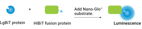
Studying proteins in their natural context is one of the biggest challenges in biology. From tumor suppressors to growth factors, some of the most clinically-relevant proteins are also the hardest to study. One common strategy is protein overexpression — boosting levels so ...
It was huge news earlier this year: the first patient in the world, an infant, was successfully treated with a CRISPR gene editing therapy personalized to his genetic mutation. “Baby KJ” was diagnosed with a rare and dangerous metabolic disease shortly after his birth. Within a ...
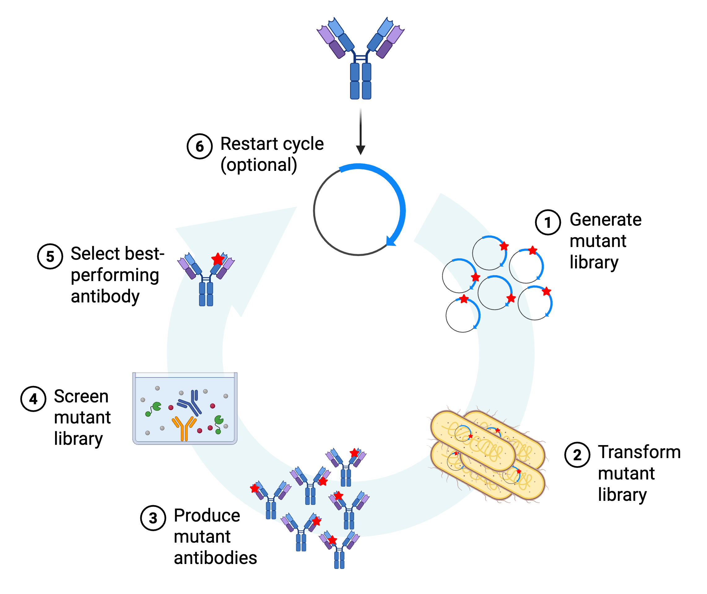
It takes a village! While you usually hear this phrase when discussing raising children, it can also be applied to research. Sharing knowledge and tools is the best way to help propel research forward and have the biggest impact. We should know — Addgene has built its foundation ...
Every few months we highlight some of the new plasmids, antibodies, viral preps, and more in the repository through our Hot Plasmids articles.
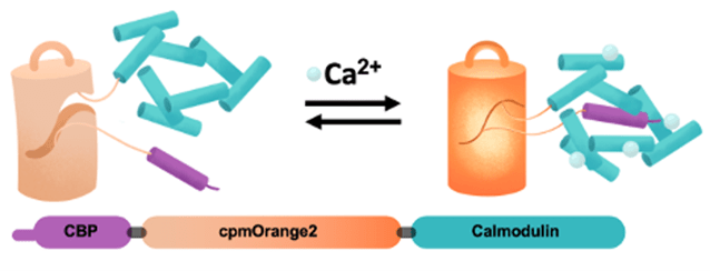
This post was written by Abhi Aggarwal, from University of Calgary. Over the past few decades, genetically encoded calcium indicators (GECIs) have become a vital tool in neuroscience research. These fluorescent proteins light up in response to calcium, which is more than just ...
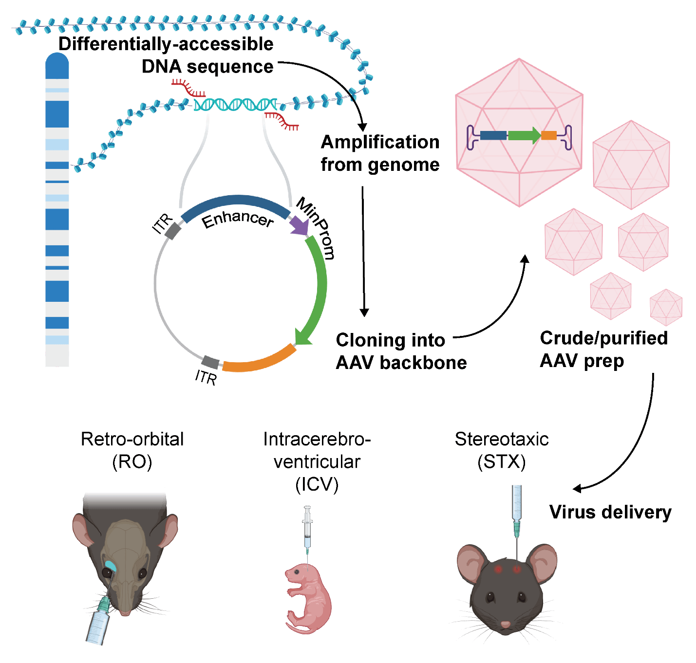
This post was written by Alyssa Shepard and Angelo Nicolaci, a PhD student at Moffitt Cancer Center. Engineering usually calls to mind building things, lots of math, and maybe heavy machinery. But not all engineering is at a large scale — some is done on much smaller equipment ...
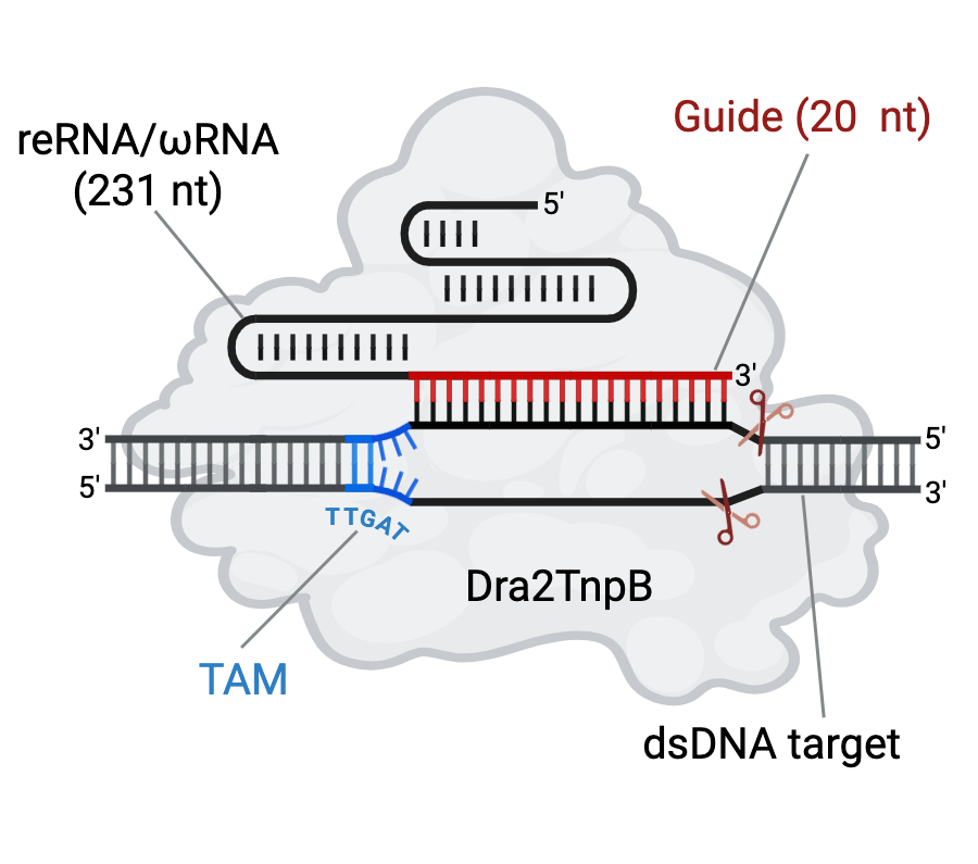
This blog post was written by Dr. Kutubuddin Molla, investigator at ICAR-Central National Rice Research Institute. When it comes to genome editing, CRISPR is a name that resonates with nearly every biologist, academic, and researcher. Among the most well-known CRISPR-associated ...