Every few months we highlight a subset of the new plasmids, antibodies, and viral preps in the repository through our hot plasmids articles. Think we missed something hot? You can pitch a Hot Plasmids section (antibodies and viral preps welcome!) here.

Here's what you'll find in this post:
- Improved voltage indicator: JEDI-2P
- Novel class of light-gated potassium channelrhodopsins
- Myc-tag antibody anti-c-Myc [9E10] now available!
Improved voltage indicator: JEDI-2P
by: Brian O'Neill
The François St-Pierre lab and collaborators have successfully developed an improved voltage indicator (GEVI) called JEDI-2P. Using a custom, high-throughput screening platform to apply saturating mutagenesis on 21 positions in the protein sequence of the predecessor GEVIs (called ASAP 1 and 2), they found an indicator that is optimized for 2-photon imaging of voltages. This includes monitoring of fast bursts, as well as slow up/down voltage states in deep tissue, in vivo. Two of their constructs are currently available as Cre-dependent, EF1ɑ-driven viral vectors (AAV1) - and you can also find several other constructs in plasmid form.
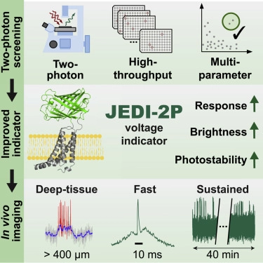 |
| Fig. 1: Summary of the approach and outcomes of the screen for improved genetically-encoded voltage indicators (GEVIs) based on the ASAP sensors. The resultant JEDI-2P sensors overcome many challenges in 2-photon in-vivo imaging of voltage fluctuations. Image credit: Liu Z, et al. Cell. 2022. |
Find JEDI-2p viral preps here (and here!)
Find JEDI-2p plasmids here!
Liu, Zhuohe et al. Sustained deep-tissue voltage recording using a fast indicator evolved for two-photon microscopy. Cell, 18, 3408 - 3425.e29. (2022). https://doi.org/10.1016/j.cell.2022.07.013.
Novel class of light-gated potassium channelrhodopsins
by: Mike Lacy
New work from the labs of Mingshan Xue and John Spudich identifies a novel class of light-gated potassium channelrhodopsins, named Kalium channelrhodopsins (KCRs), from a fungus-like protist Hyphochytrium catenoides (Govorunova et al. 2022). The authors show that HcKCR1 and HcKCR2 have high selectivity for K+ over Na+ and rapid photocurrent kinetics. They demonstrate the use of HcKCR1 as an optogenetic silencer in experiments with mouse cortical neurons, far out-performing previous attempts to engineer potassium-selective channelrhodopsins. HcKCR1 has a maximum absorption of 540 nm and two-photon excitation around 1,040 nm, has larger photocurrents, and is more selective for K+ than HcKCR2, making it more suitable for in vivo optogenetic use.
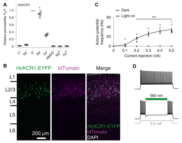 |
| Fig. 2: A) HcKCR1 is highly selective for K+ over other Na+ and other cations. B) Cortical slice of HcKCR1-EYFP and tdTomato expressed layer 2/3 neurons in mouse. C) Action potential frequencies with (magenta) and without (black) photoactivation of HcKCR1 in mouse neurons. D) Membrane voltage traces of action potentials in a HcKCR1-expressing mouse neuron without (top) and with (bottom) photoactivation of HcKCR1. Images adapted from Govorunova et al. 2022. |
HcKCRs represent a long-sought tool for researchers to control K+ gradients and can be used as a potent optogenetic silencer. The current work also proposes promising avenues for future engineering to shift the spectra and improve the performance of HcKCRs.
Govorunova EG, et al. Kalium channelrhodopsins are natural light-gated potassium channels that mediate optogenetic inhibition. Nat Neurosci 25, 967–974 (2022). https://doi.org/10.1038/s41593-022-01094-6. (This work was also shared as a preprint: https://doi.org/10.1101/2021.09.17.460684.)
Myc-tag antibody anti-c-Myc [9E10] now available!
by: Ashley Waldron
The majority of Addgene's more than 50 antibodies target neuroscience-related proteins, but we recently added several broadly useful antibodies for detecting protein tags.
And when you think of protein tags, what are the first tags that come to mind? I’m willing to guess that the Myc-tag is one of them (and not just because of the title!) The Myc-tag is a string of 10 amino acids, EQKLISEEDL, that can be incorporated into your protein of interest and recognized by reliable antibodies. It is derived from the C-terminal region of the human proto-oncogene c-Myc. Since the generation of the first anti-c-Myc [9E10] (Evan, et al., 1985), the Myc tag has become one of the most commonly used protein tags - a quick search for ‘Myc tag’ in the Addgene catalog pulled up over 13,000 plasmids! Our recombinant anti-c-Myc [9E10] (isotype IgG2a) antibody is currently recommended for use in western blot, but we are actively testing it in other applications! If you try it out in a new assay, let us know. We’d love to hear from you!
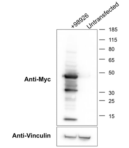 |
| Fig. 3: Image of Addgene’s western blot for the myc-tagged protein GFP-smFP-myc expressed from Plasmid 98926. See the Applications section on the Anti-c-Myc [9E10] page for more details. Image credit: Addgene. |
Find anti-c-Myc antibody here!
Evan, G. I., Lewis, G. K., Ramsay, G., & Bishop, J. M. (1985). Isolation of monoclonal antibodies specific for human c-myc proto-oncogene product. Molecular and Cellular Biology, 5(12), 3610–3616. doi: https://doi.org/10.1128/mcb.5.12.3610-3616.1985.

Topics: Hot Plasmids


Leave a Comment