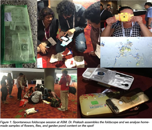
When you think about going to a scientific conference, you may think about sitting amongst a sea of chairs listening to talks all day. But nope, not at the American Society for Microbiology 2018 Microbe meeting. Soon after I arrived, I was looking through a paper-based, affordable, and portable microscope called the ‘foldscope’ (Dr. Prakash, Stanford University). Right there on the floor outside the lecture hall.
It was wonderful to see the ad-hoc ‘sciencing’ when Dr. Prakash (twitter: @PrakashLab #foldscope) had taken out a few foldscopes and held an improv workshop. These paper microscopes work as conventional microscopes and give a similar optical quality (magnification of 140X and 2 micron resolution).
I have worked many hours in a dark room with a confocal microscope, yet I was once again fascinated by the world that reveals itself when looking down a paper thin and very portable microscope. Less fluorescence admittedly, but still so beautiful! Enabling the scientist in everyone - everywhere!
From ‘looking at’ to ‘listening to’ microbes
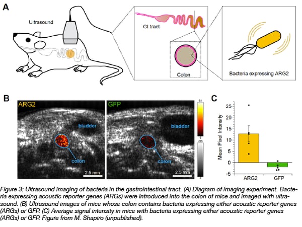
But seeing microbes through a microscope is not the only way to visualize them. You can also “hear” them. Addgene depositor Dr. Mikhail Shapiro from Caltech presented an exciting way of listening to microbes by using non-invasive methods for cellular imaging with ultrasound. In his research he applies naturally occurring bacterial “molecular buoyancy devices” (also known as gas vesicles) as acoustic biomolecule-based dyes. This technology is widely applicable as bacterial expression of dyes allows dye visualization in vivo using ultrasound (for an example see the figure below). In addition to low costs, deep tissue penetration, and high spatial resolution are benefits of this technique. It is even possible to multiplex ultrasound dyes because these dyes “collapse” at different ultrasound frequencies. Read more about ‘Using ultrasound to image bacteria in vivo: acoustic reporter genes’ on our blog.
Creative approaches to presenting the microbial world
After learning about using sound to visualize bacteria in the lab, I was excited to learn about creative approaches to visualize bacteria in an educational setting. The Symposium on ‘Creative Approaches to Presenting the Microbial World’ showcased complex connections and ideas in various visualized approaches from hand-drawn comics (Dr. Sanja Saftic) to educational gifs created by the Amoeba Sisters. Here’s one example of how they “bridge the jargon gap using gifs”:
The take home message was that being creative and telling a story makes scientific topics both interesting and approachable!
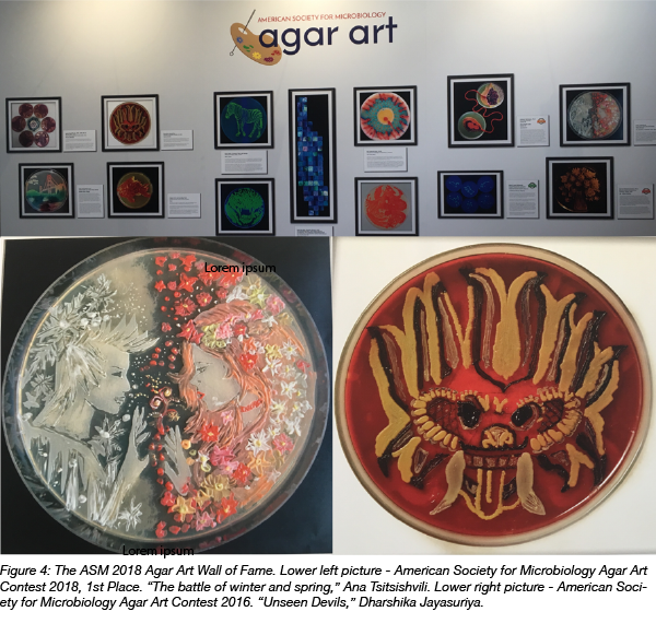
The grand finale of visual impressions was delivered by the ASM Agar Art Winners! It is absolutely fascinating to see the living art designed by creative minds with a (I assume) very steady hand. By connecting art and science, a fascinating interface is created that produces loads of inspiration for the current and the next generation of microbiologists.
If you got inspired to produce your own agar art have a look at our Microbiology and Fluorescent Protein Resources and get started!
I learned a lot about creative ways to do science communication and outreach - if you'd like to learn more about Science communication check out these SciComm blog posts.
References
1. Bourdeau, Raymond W., et al. "Acoustic reporter genes for noninvasive imaging of microorganisms in mammalian hosts." Nature 553.7686 (2018): 86.PubMed PMID: 29300010. PubMed Central PMCID: PMC5920530.
Resources on the Addgene Blog
- Read Our Post on CRISPR Methods for Bacterial Genome Engineering
- Learn about Science Communication using Twitter
Resources on Addgene.org
Topics: Microbiology, Other


Leave a Comment