


Every few months we highlight some of the new plasmids, antibodies, viral preps, and more in the repository through our Hot Plasmids articles. This month, we’ve got lots of exciting materials to talk about, from the latest prime editors and new CRISPR tools to antibodies and a viral astrocyte tracer for neuroscience research. There’s so much to share, let’s dive in!
 Read on to find:
Prime editors get an AI-boosted redesign
By Emily P. Bentley
Prime editing is a "search-and-replace" genome editing technique that combines Cas9, a reverse transcriptase (RT), and a pegRNA that specifies the genomic target and edit in a single sequence. Over the years, prime editors have been through many generations of optimization. But every mutation involves a tradeoff, and in 2026, the David Liu Lab showed that recent prime editors had unstable RT domains — the price of their improved efficiency (Tao et al., 2026). That instability reduced protein expression and could offset efficiency gains.
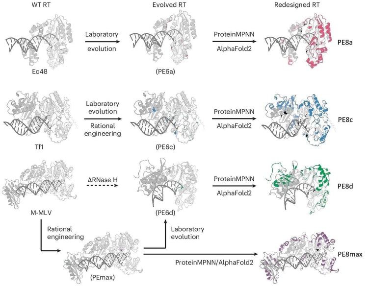
To escape these constraints, the team used ProteinMPNN, a protein sequence design tool, to vary the sequences of PEmax, PE6a, PE6c, and PE6d, aiming to improve their thermodynamic stability while restricting the program from altering catalytic or highly conserved residues. Hundreds of redesigned prime editors were validated with AlphaFold and screened for editing efficiency, including in a therapeutically relevant lipid nanoparticle (LNP) delivery system.
The best performing editors were designated PE8max, PE8a, PE8c, and PE8d, according to their predecessors (Figure 1). Each has unique benefits. The authors recommend PE8c for most workflows, though PE8d and PE8max can achieve higher editing efficiencies at some sites. PE8a is the smallest editor, at 5.6 kb, and so works best for size-constrained contexts. Read our updated blog post for more advice on selecting the best PE8 variant for your next experiment.
 |
|
Figure 1: AI-guided development of PE8 series reverse transcriptase (RT) domains, with changes shown in color against the original sequence in gray. Image from Tao et al., 2026, under a CC BY-NC-ND 4.0 license. |
Tao, Y. A., et al. (2026). AI-guided redesign of laboratory-evolved reverse transcriptases enhances prime editing. Nature Biotechnology, 1–13. doi: https://doi.org/10.1038/s41587-026-03149-6.
A hot Cas summer for shredding DNA
By Alyssa Shepard
Cas enzymes are well-known for their ability to chop DNA. Generally, they are put into practice to precisely eliminate or silence one or a handful of genes at a time. But what if we could go bigger?
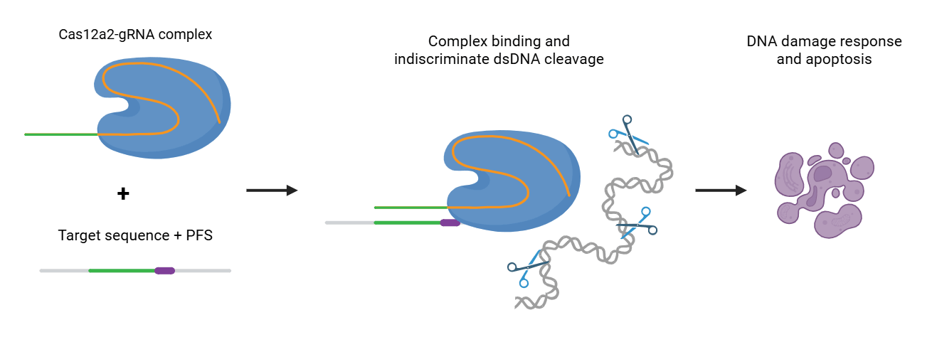
A newly discovered version of Cas12a, dubbed Cas12a2, is tearing up DNA, one cell at a time. Instead of a highly localized DNA cleavage mechanism, Cas12a2 "unleashes indiscriminate double-stranded DNase (dsDNase) activity" when it binds its target RNA (Scholz, Thompson, Crosby et al., 2026). This leads to the DNA in a cell effectively being shredded, which naturally causes the cell to panic and put their DNA damage responses into overdrive, eventually triggering cell cycle arrest and apoptosis. In this way, a targeted Cas12a2 enzyme can lead to the downfall of a population of cells.
 |
|
Figure 2: Overview of Cas12a2 activity. The gRNA in the Cas12a2-gRNA complex contains a targeting protospacer specific to the cell population of interest. This binds with the target RNA sequence, which includes the protospacer flanking sequence (PFS). Upon binding to the target, Cas12a2 begins double-stranded DNA (dsDNA) cleavage, leading to increased DNA damage response and apoptosis. Image created with BioRender.com. |
Two groups have now put this enzyme into action in mammalian cells — led by Yang Liu and Jennifer Doudna. The Liu group tested Cas12a2 in yeast and human cell lines and found that they could eliminate virally-infected cells, destroy cells harboring oncogenic mutations, and enrich for gene-edited cell populations (Scholz, Thompson, Crosby et al., 2026). Likewise, the Doudna group focused on targeting cancerous populations. They found similar results, using Cas12a2 to selectively target and degrade cell populations with overexpressed oncogenes, neojunctions, and specific mutations in TP53 (Zeng et al., 2026). They were even able to show a reduction in tumor growth in mice by targeting TP53 and MYC mutations. Both labs utilized the same Cas12a2 enzyme (SuCas12a2, from Sulfuricurvum sp. PC08-66), with different selection markers for diverse experimental set ups. These Cas12a2 plasmids have been distributed to dozens of labs already, so get yours and get ready to shred some DNA!
Find the Cas12a2 plasmids from the Liu and Doudna Labs!
Scholz, P., Thompson, J., Crosby, K. T., et al. (2026). RNA-triggered cell killing with CRISPR–Cas12a2. Nature, 655(8121), 230–239. https://doi.org/10.1038/s41586-026-10466-y.
Zeng, J., et al. (2026). Targeting cancer-specific mutations with RNA-triggered chromatin shredding. Nature, 656(8126), 199–206. https://doi.org/10.1038/s41586-026-10738-7.
Mapping bivalent chromatin with CoCUT&Tag
By Mike Lacy
There are many ways to map chromatin modifications or proteins across the genome, from classic methods like ChIP to newer workflows like CUT&RUN or CUT&Tag. But understanding how multiple features coexist at the same site is still a challenge, and researchers often rely on correlations between independent experiments. Now, the Derek Janssens Lab has developed CoCUT&Tag (co-occupancy Cleavage Under Targets and Tagmentation) which overcomes key limitations to directly map pairs of chromatin factors on the same DNA (Sun et al., 2026).
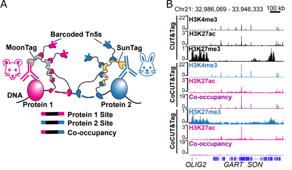
In CUT&Tag, antibodies direct a DNA transposase to a chromatin feature, then the transposase cleaves the local DNA while tagging the fragments with adaptors ("tagmentation"), to be amplified and identified through sequencing. In CoCUT&Tag, secondary antibodies are replaced by arrays of epitope tags (SunTag and MoonTag), to incorporate unique barcoded adapters at any pair of targets recognized by the primary antibodies (Figure 3).
 |
|
Figure 3: Measuring chromatin co-occupancy of multiple target features with CoCUT&Tag. A) Schematic of CoCUT&Tag. B) Representative genomic tracks for CUT&Tag on three histone modifications. Co-occupancy signal is detected at loci marked by H3K4me3 and H3K27ac, but not over the loci individually marked by H3K27me3 and H3K27ac. Adapted from Sun et al. (2026) under a CC BY license. |
Individually, the new constructs performed similarly or better than conventional CUT&Tag, but their main advantage is when used in combination to map bivalent chromatin. coCUT&Tag enables single-cell measurements as well as bulk profiling with much less input material than would be required for co-ChIP (<50,000 cells instead of millions). Whether verifying past correlations or asking brand new questions, this approach will let you chart a new map — even in familiar territory.
Sun, W., Baird, E., Ashbaugh, H. J., Eiken, A. P., Janssens. D. H. (2026). CoCUT&Tag maps linked chromatin states at single-molecule, single-cell resolution. bioRxiv 2026.04.27.721191; doi: https://doi.org/10.64898/2026.04.27.721191.
Speeding up worm transgenesis with one simple plasmid
By Isabella Lima
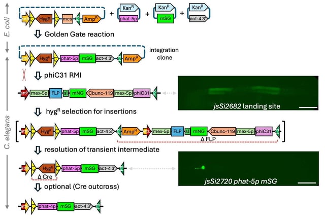
C. elegans researchers are always looking for new and improved methods for transgenesis that minimize work and time required. Now, thanks to the Michael Nonet Lab, single copy transgenic lines can be created in 6–8 days without crossing, only injection. This work uses a novel recombination-mediated insertion (RMI) landing site that contains the recombinase cassette in the worm genome (Beck & Nonet, 2025). This makes it possible to incorporate a transgene with just a single target vector, eliminating the need for a separate driver and reporter (Figure 4). Injection into a single worm has shown to yield multiple independent insertions, increasing the likelihood of successful integration of the transgene.
 |
|
Figure 4: Overview of the single-component RMI plasmids and workflow. An integration clone is constructed using Golden Gate cloning, then is injected into the RMI landing site strain jsSi2682. The resulting strain, jsSi2720 phat-5p mSG shows strong fluorescent signal in pharyngeal gland cells. Image adapted from Beck & Nonet (2025) under a CC BY 4.0 license. |
The authors have deposited two integration vectors, for selection with either hygromycin or neomycin. The desired DNA is inserted into the vector (by Golden Gate cloning) and then injected into the worms. After one generation, the antibiotic can be added to efficiently select for worms that have successfully integrated the transgene. They demonstrate the value of this method by generating worms expressing over a dozen fluorescent reporters in pharyngeal gland cells to study promoter function. These new plasmids and the RMI technique promise to make transgenesis easier, faster, and more reliable.
Plus, check out the Nonet Lab’s other recent publication on integrating bipartite expression transgenes, including even more RMI targeting vectors!
Find the single-component RMI plasmids here!
Beck AA, Nonet ML. 2025. A single component landing site for efficient transgenesis using recombination-mediated insertion. microPublication Biology. doi: https://doi.org/10.17912/micropub.biology.001784.
Fishing for bacterial immune triggers with the Phage ORFeome Library
By Emily P. Bentley
The bacterial immune system isn’t limited to CRISPR: over 100 antiphage systems are known to sense bacteriophage infection and mobilize a cellular response. These systems are diverse, and it is challenging to predict their triggers.
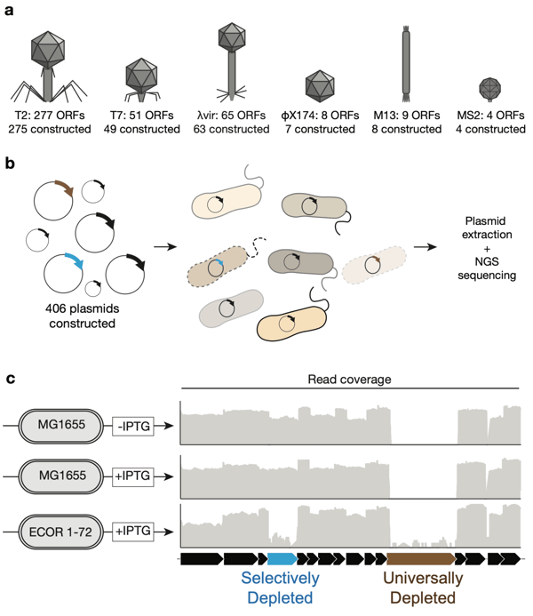
That’s why the Aaron Whiteley Lab assembled the Phage ORFeome Library, a pooled library of every open reading frame from phages T2, T7, λ, MS2, φX174, and M13 — missing only eight ORFs that were seemingly toxic even at uninduced levels (Nagy et al., 2026). After excluding vectors that replicated poorly in a K-12 lab strain of E. coli that lacks most antiphage systems, this library was transformed into 72 "wild" E. coli strains. Most antiphage responses kill the infected bacterium as well as the phage, so the team could screen for activation of these systems simply by observing which plasmids were lost from the population over time (Figure 5).
 |
|
Figure 5: A genetic screen to identify candidate phage triggers. A) Plasmids expressing every phage open reading frame (ORF) predicted to encode a protein were constructed. Phage ORFs from the genomes of dsDNA phages (T2, T7, λ), ssDNA phages (φX174, M13), and a ssRNA phage (MS2) were expressed under the control of an IPTG-inducible promoter. Plasmids for 406 out of a total of 414 ORFs could be constructed. B) Individual plasmids were pooled and transformed into bacterial strains of interest. By transforming K-12 E. coli (strain MG1655), proteins that are generally growth inhibitory could be identified. By transforming diverse E. coli from the ECOR collection, proteins that are growth inhibitory to specific strains could be identified. C) Transformants were recovered on solid agar with or without 50 µM IPTG. Plasmids were then extracted from the pooled population, sequenced, and reads mapped to phage genomes. Representative data shows read depth on a log scale for reads aligned to a portion of the phage λ genome. Figure reproduced from Nagy et al., 2025 under a CC-BY-NC-ND 4.0 license. |
Their screen yielded over 100 pairs of candidate antiphage triggers and the E. coli strains that are sensitive to them, including both known and previously undescribed antiphage systems. The team was able to identify the activator of a known antiphage system — a conserved fold in phage capsid proteins — and characterize a novel system that senses virion tail fiber proteins. With such a diverse library, there’s plenty more for you to discover!
Find the Phage ORFeome Library here!
Nagy, T. A., Gersabeck, G. W., Conte, A. N., & Whiteley, A. T. (2026). A phage protein screen identifies triggers of the bacterial innate immune system. Nature Microbiology, 11(2), 597–609. https://doi.org/10.1038/s41564-025-02239-6. (Preprint: 2025, bioRxiv 2025.07.02.662641. https://doi.org/10.1101/2025.07.02.662641.)
Mapping the astrocyte connectome with a gap junction-targeted biotin ligase
By Brian O'Neill
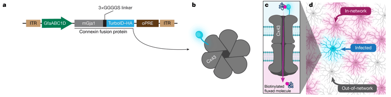
When we talk about brain connectome mapping, you probably think neurons — but the connections between other cell types like astrocytes are also complex and important for brain function. The labs of Moses Chao and Shane Liddelow recently built an astrocyte network tracer to reveal these connections (Cooper et al., 2026). By fusing TurboID with the gap junction protein connexin 43 (Cx43/GJA1), they could tag proteins that pass through the gap junction channel from one astrocyte to its neighbors (Figure 6).
 |
|
Figure 6: Astrocyte network tracer. A) Schematic of the astrocyte network tracer construct. B–C) One Cx43 subunit fused with TurboID-HA (blue) is incorporated into a gap junction channel alongside untagged endogenous subunits. As molecules pass through the channel, they are first biotinylated (magenta) by TurboID at the channel vestibule. D) Labeling with streptavidin and anti-HA then imaging identifies in-network cells (HA-/streptavidin+, magenta), initial infected cell (HA+/streptavidin+, blue), and out-of-network cells (HA-/streptavidin-, gray). Image reproduced from Cooper et al. (2026) under a CC BY-NC-ND 4.0 license. |
Using mice injected with AAV5 to express the astrocyte network tracer, followed by whole-brain tissue clearing and imaging of both the tagged AAV transgene (only present in "starter cells") and the biotinylated proteins (present in any "in-network" cells), they showed that astrocytes form long-range networks. Amazingly, these networks have distinct shapes depending on the brain region of the starter astrocytes, and the networks do not necessarily follow the long-range paths of neurons from that brain region. These long-range astrocyte networks are structurally plastic and may enable novel communication between brain regions.
This tool will be very useful in studying the astrocytic proteome and the network’s role in distributing these molecules. Using the tracer to map the networks, combined with other techniques to study their function, promises to reveal many new findings.
Find the astrocyte network tracer plasmid here!
Cooper, M. L., et al. (2026). Astrocytes connect specific brain regions through plastic networks. Nature, 655(8121), 183–191. https://doi.org/10.1038/s41586-026-10426-6.
New antibodies for studying axon guidance
By Ashley Waldron
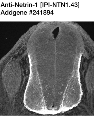
Netrin and DCC are well-known proteins involved in axon guidance. Classically, Netrin-1 interacts with DCC to direct commissural axon navigation during spinal cord development. But of course, the full picture is much more complex. For example, netrins can exert opposing actions depending on the cellular context, and there are still members of the DCC protein family that have yet to be fully characterized. Researchers have also revealed new associations between these proteins and human diseases, emphasizing the importance of untangling the complex functions of these proteins. To support such efforts, the Institute for Protein Innovation has developed a new collection of highly specific, community-validated antibodies against DCC, Netrin-1, and IGDCC3 (PUNC). These chimeric recombinant antibodies have been tested for flow cytometry, immunocytochemistry, and immunohistochemistry. Full details and validation data across applications are available on each product page, so you can start your experiments with confidence.
 |
|
Figure 7: Immunohistochemistry with Netrin-1 antibody from IPI. Rat embryo spinal cord sections (30 µm) were fixed and labeled with Anti-Netrin-1 [IPI-NTN1.43] (Addgene #241894), showing clear labeling of the floor plate. Image from Butler & Mercado-Ayon 2025, Addgene Report, https://doi.org/10.57733/addgene.amfit7. |
Find Netrin, DCC, and IGDCC3 (PUNC) antibodies here!
That's all for now! If you've tried these tools in your lab, let us know what you think in the comments below.
Topics: Hot Plasmids



Leave a Comment