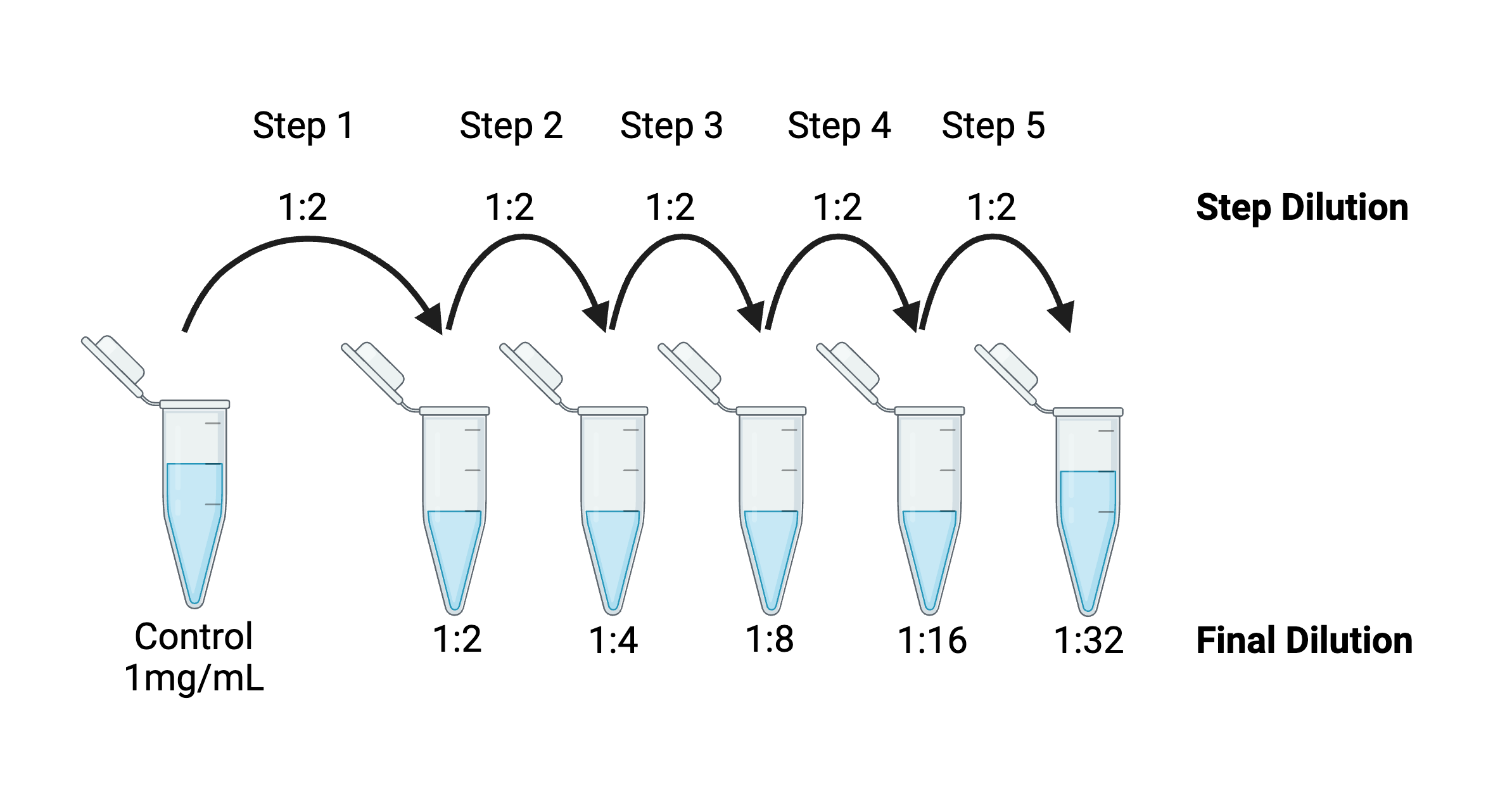
What do a viral vector production facility, food allergy testing lab, and the grad student down the hall from you have in common? All of them rely on standard curves in their day-to-day work. Indeed, viral vector production facilities frequently use qPCR with a standard curve to ...
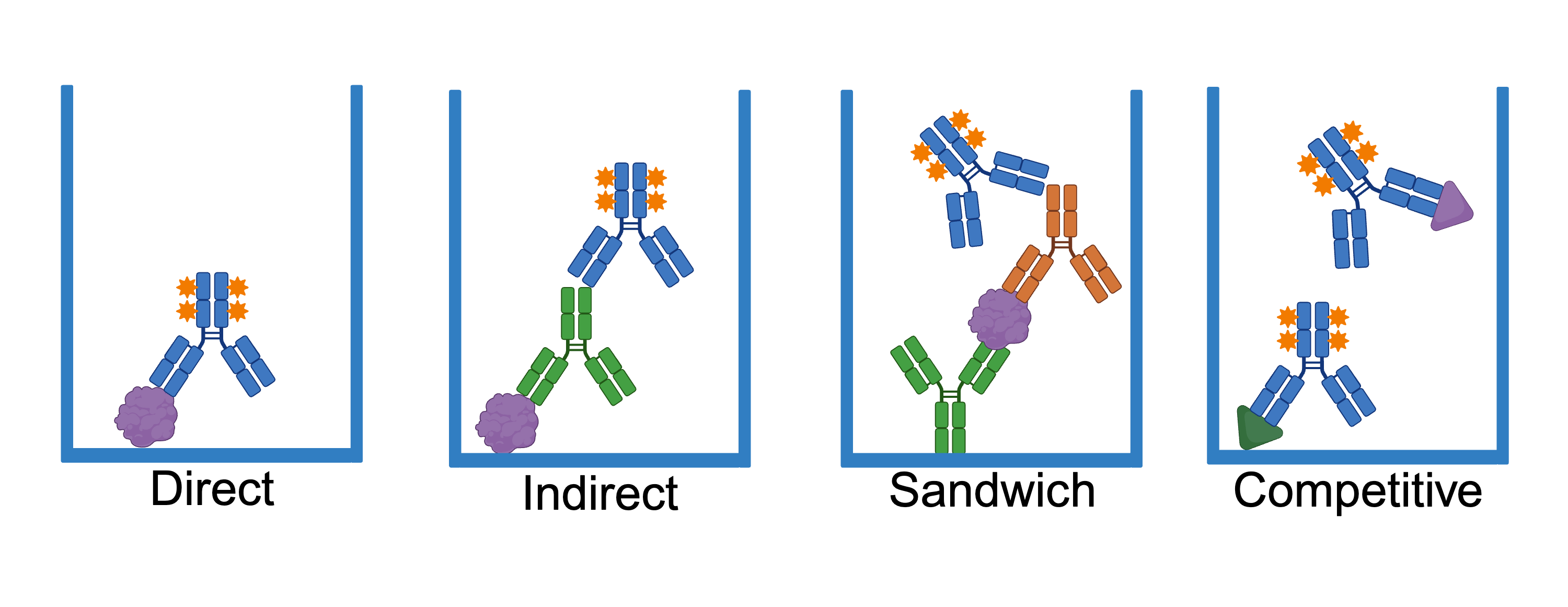
An enzyme-linked immunosorbent assay (ELISA) is a versatile method used to quantify the level of target antigen in a sample. While Engvall et al. originally developed the ELISA assay to measure antibody levels, scientists have since adapted it for a host of different proteins ...
In a world where so much is out of your hands, it’s helpful to focus on something controllable, like experiments (and their controls!). This blog post will discuss the ins and outs of controls for biological experiments, starting with general controls and then moving on to ...
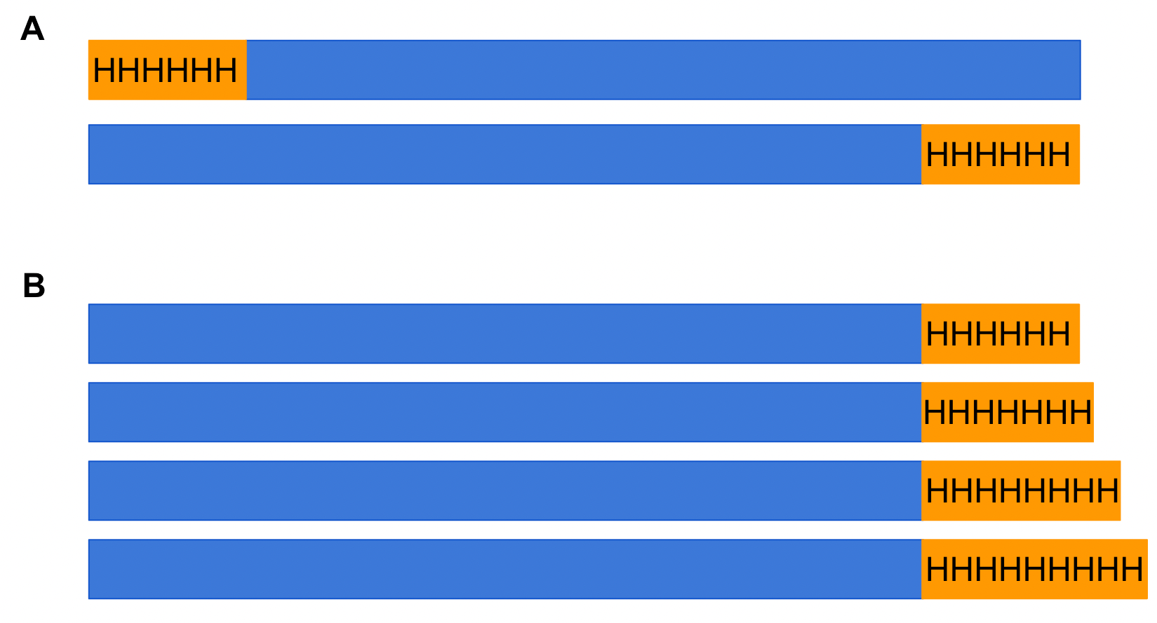
You are a scientist looking to determine how Protein A and Protein B interact. You read extensive research on the two proteins and come up with a great experimental plan that requires indirect staining of both targets in your specimen. You scour the literature and find an ideal ...
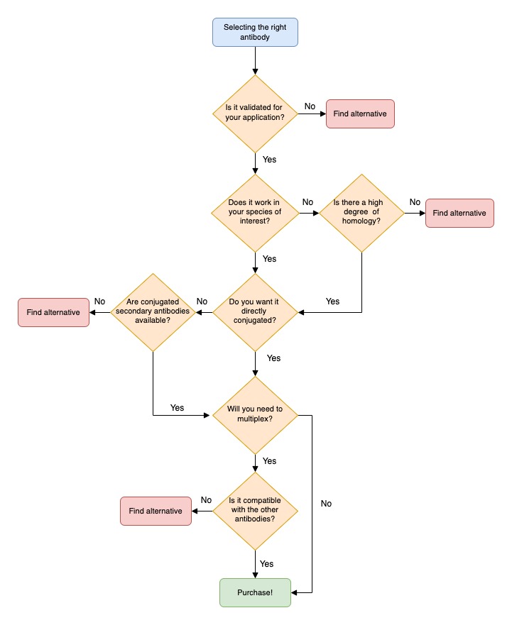
If you are lucky in lab life, you will have a plethora of antibody options for your experiment (all well validated for your application, of course!) When the stars are aligned and the lab gods are smiling down at you, you may wonder which antibody should I pick? Do I go for the ...
Much of today's biological research requires a close examination of specific proteins within a system. This can be pretty complicated given that a single cell has tens of thousands of proteins functioning in a variety of ways. How do scientists focus on the activity or function ...
Picture this: you’ve been assigned an exciting new project aimed at understanding how a critical cellular pathway is regulated. You’ve read all the background papers you could get your hands on, formulated a hypothesis, and planned out your key experiments. Unsurprisingly, many ...
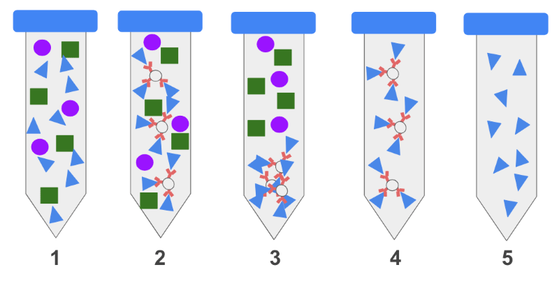
Immunoprecipitation (IP) uses immobilized antibodies, or immunoglobulins, to isolate a specific protein out of a complex mix. Using this technique, users can look for the presence or absence of a protein, determine if a protein is up or downregulated, examine a protein’s ...


-min.jpg)