I often wonder about the gut-brain axis (admittedly for self-serving reasons – I want to understand my obsession with the combination of chocolate and peanut butter), but it’s an undeniably difficult connection to study on a molecular level. The brain is constantly influenced by signals it receives from the entire body, making it difficult to probe due to the lack of tools to continuously monitor it. How to address this? Through the pairing of materials scientists (Anikeeva lab) with gut-brain biologists (Bohórquez lab), who together developed multifunctional fibers, embedded, and validated simultaneously in the brain and the intestines of mice. The result? A very cool tool to advance our knowledge of brain-visceral organ signaling.
The problem
Previously, there were very few tools available for monitoring how both the brain and visceral organs interact during various behaviors. Either tissue samples were harvested after the behavior, or the animals being monitored were physically hooked up to invasive, cumbersome systems during experiments – neither ideal for the intrepid researcher. There existed, therefore, a need for an adaptable tool that could continuously monitor multiple systems. Ideally this tool would be wireless, able to monitor outcomes of interest, and stimulate or manipulate responses through neurological means like electric stimulation, drug administration, and optogenetics. Enter neural fibers!
The neural fiber tool
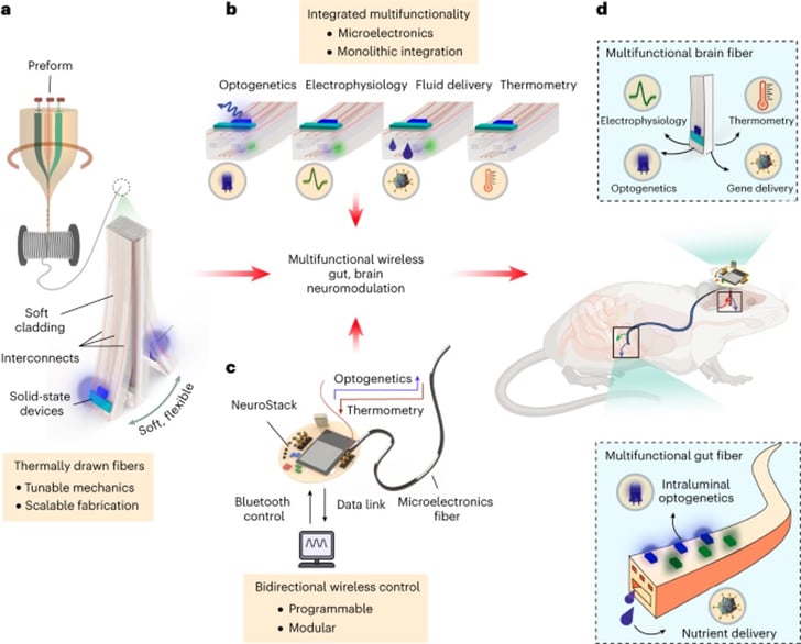
The neural fibers developed by the Anikeeva lab are microelectronic fibers made from thermoplastic polymers that can work with all the bells and whistles. The fibers are made from a process called thermal drawing, essentially where blocks of polymers are heated up and pulled to a desired thickness. The fibers are drawn with microelectronics included in them for downstream control – this is important later! Next, additional components are added - the ‘bells and whistles’ – including microscale light emitting diodes, thermal sensors, and microfluidic channels. These features enable the fibers to emit light for optogenetic purposes, measure temperature, study electrophysiology, and deliver small molecules.
Fun fact: A 1 cm block of polymer can generate many meters of fibers!
|
|
Fig. 1 - Overview of the neural and gut fiber system (adapted from Sahasrabudhe et al., 2023). |
To develop these fibers, the labs looked at previous implementation of optogenetic tools, calcium sensors, and other common neuroscience tools into the brains of model organisms, including the Anikeeva lab’s work on a similar neural fiber (Park et al., 2019). This provided an engineering road map of sorts, giving an idea of the stiffness the fibers would need to be, as well as other features necessary for implantation in brain tissue.
The gut, on the other hand, was the wild west for fiber implants. Due to the soft nature of the intestines and the requirement for digestive motility to not be disturbed, the brain fibers could not be directly repurposed for gut implantation. The fibers they ended up generating were 10-15 times more flexible than the brain fibers and importantly distortion of the fibers didn’t affect their function (the gut twists and contorts during the digestion of food, while brain tissue is relatively static).
To operate both types of implanted fibers simultaneously, a mini-Bluetooth system named NeuroStack was used (Orguc et al., 2020). Using a programmable microcontroller, NeuroStack coordinates optical stimulation of its associated fibers across multiple channels and can also record tissue temperature proximal to the implanted fibers. As important as its capabilities are the practical considerations of actually using it - the device has a rechargeable battery, is relatively small, and can be detached easily whenever necessary.
This is all an impressive engineering feat, but you’re probably wondering “did it actually work”? The authors first tested that the system could induce stimulation in target tissues of a live animal. They started by delivering an AAV construct expressing channelrhodopsin 2 (ChR2), a blue light sensitive cation channel, fused to mCherry to the brain tissue. They performed optical stimulation using NeuroStack and the fibers as previously described, then measured electrophysiological potential afterwards. This measurement trended with expression of the opsin in the target neurons, validating the functionality of these inputs and outputs for the system. Success! They also observed reliable optically evoked neuron activity for at least several months after implantation of fibers, indicating the system was indeed stable for continuous monitoring. On the gut side of things, the authors optigenetically and chemically excited a class of enteroendocrine cells within the intestines known as neuropods. They observed increased vagus nerve firing in response to both of these stimulations. The best part? The chemical stimulation was sucrose…..sounds like the gateway to chocolate and peanut butter to me.
What’s on the fiber horizon?
The brain interfaces with the entire body, so the obvious next applications are to expand the tool to different brain-organ axes. Alternatively, the system could be used to deliver or measure new variables. The sky is really the limit for this emerging technology!
Resources and References
References
Orguc, S., Sands, J., Sahasrabudhe, A., Anikeeva, P., & Chandrakasan, A. P. (2020). Modular Optoelectronic System for Wireless, Programmable Neuromodulation During Free Behavior. 2020 42nd Annual International Conference of the IEEE Engineering in Medicine & Biology Society (EMBC), 4322–4325. https://doi.org/10.1109/EMBC44109.2020.9175600
Park, S., Loke, G., Fink, Y., & Anikeeva, P. (2019). Flexible fiber-based optoelectronics for neural interfaces. Chemical Society Reviews, 48(6), 1826–1852. https://doi.org/10.1039/C8CS00710A
Sahasrabudhe, A., Rupprecht, L. E., Orguc, S., Khudiyev, T., Tanaka, T., Sands, J., Zhu, W., Tabet, A., Manthey, M., Allen, H., Loke, G., Antonini, M.-J., Rosenfeld, D., Park, J., Garwood, I. C., Yan, W., Niroui, F., Fink, Y., Chandrakasan, A., … Anikeeva, P. (2023). Multifunctional microelectronic fibers enable wireless modulation of gut and brain neural circuits. Nature Biotechnology, 1–13. https://doi.org/10.1038/s41587-023-01833-5
Resources on Addgene.org
More resources on the Addgene blog
Topics: Neuroscience


Leave a Comment