This post was contributed by guest blogger Tessa Cressey.
The highly pathogenic Ebola virus belongs to the group of nonsegmented negative sense RNA viruses, along with other viruses that cause disease in humans such as measles, mumps, and rabies. Research on Ebola virus has been limited, in part, due to the necessity for working with this virus under the highest biosafety level conditions, BSL-4. In this regard, minigenome systems, such as the one developed for Zaire ebola virus (EBOV) are extremely useful, allowing researchers to study aspects of the EBOV replication cycle under BSL-2 conditions (4).
What is a minigenome?
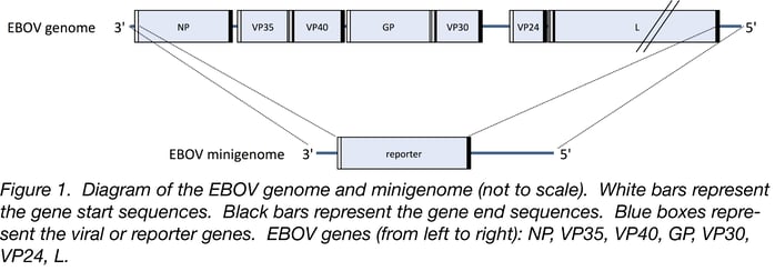
 The EBOV genome is a nonsegmented, negative sense RNA strand approximately 19 kb in length. It contains short promoter sequences at its 3’ and 5’ ends that are crucial cis-acting signals for genome replication and viral gene transcription (6). The EBOV genome contains seven viral genes, each flanked by a transcription start signal sequence (gene start) and a transcription termination/polyadenylation signal sequence (gene end).
A minigenome is a shortened version of the viral genome. It contains the 3’ and 5’ ends of the genome which are required for replication and transcription (Figure 1). All viral genes are removed and replaced by a single (non-viral) reporter gene (e.g. firefly luciferase or eGFP) flanked by the EBOV-specific gene start and gene end sequences. Thus, the minigenome contains all cis-acting signals necessary to direct minigenome replication and reporter gene transcription by the EBOV polymerase (reviewed in 2, 5). The minigenome is capable of being replicated and transcribed in the cell when the appropriate viral proteins are provided in trans (see Other Important Components of EBOV Minigenome System). However, minigenomes are incapable of producing infectious virus (similar to lentiviral vectors) because they do not encode all of the necessary viral proteins. Minigenome systems have been developed for many nonsegmented negative sense RNA viruses (reviewed in 2, 3).
Other important components of the Zaire ebola virus minigenome system
Similar to other RNA viruses, EBOV brings its own RNA-dependent RNA polymerase into the infected cells. The EBOV polymerase replicates the viral genome and transcribes viral genes. Four viral proteins are required for viral genome replication and transcription: NP, VP35, VP30, and L (reviewed in 5). The RNA genome is encapsidated by the nucleoprotein, NP. NP associates with the viral polymerase, which is formed by a complex of two proteins: the large (L) catalytic protein and its cofactor VP35. VP30 is a transcription enhancement factor.
When used to study minigenomes, these four viral genes are each encoded on separate plasmids and are co-transfected into cells alongside a plasmid encoding the minigenome. These genes and the minigenome are transcribed (and the viral mRNAs are translated) by cellular machinery. NP encapsidates the negative sense, single-stranded RNA minigenome. The viral polymerase complex then recognizes the encapsidated minigenome as a template for replication and transcription, as it would for a full-length viral genome (Figure 2).
While it is technically possible to encode all four viral genes in the same plasmid (as opposed to four separate plasmids), there are a few reasons arguing against this approach. First, this would prevent the study of the proteins independently from each other (see Uses of Minigenome System). From a more technical standpoint, a plasmid with all four genes and necessary control elements for each would be relatively large. Cloning the viral genes in this plasmid would be more difficult compared to if each gene were encoded in separate (and thus smaller) plasmids. Finally, having four separate plasmids allows researchers to partially control expression levels of each gene by altering the amount transfected (see Technical Tips).
Minigenome replication and transcription
In order to replicate the minigenome, the viral polymerase initiates RNA synthesis at the 3’ end of the minigenome to produce the positive sense, genome replicative intermediate (anti-minigenome). The anti-minigenome is used as a template by the viral polymerase to synthesize additional copies of the negative sense minigenome, thus modeling genome replication. These products can be detected by Northern blot analysis.
In addition, the polymerase will use the minigenome as a template to transcribe the reporter gene (e.g. firefly luciferase). The polymerase recognizes the gene start signal and initiates transcription of the reporter gene. The polymerase will elongate the mRNA until it reaches the gene end signal, where it will terminate transcription and polyadenylate the mRNA. These mRNAs are translated by cellular host machinery (as viral mRNAs would be in the course of viral infection), producing firefly luciferase. Firefly luciferase can readily be measured to quantify (indirectly) transcription performed by the viral polymerase. Alternatively, the mRNA can be detected directly by Northern blot analysis or RT-PCR.
Uses of the minigenome system
Minigenome systems, such as the one described here, are extremely useful research tools. In addition to the reduced requirements for biocontainment precautions, these systems allow researchers to investigate viral processes in extreme detail. The mechanism of a particular observation can be easily studied as each component can be examined independently. In addition, experiments can be performed in the absence of some viral proteins (compared to viral infection). Therefore, the roles/functions of individual proteins can be studied independently of other viral proteins simplifying the interpretation of results.
This system can be used to develop an understanding of and manipulate the biology of EBOV. For instance, the roles of specific viral sequences (e.g. promoters) can be studied by changing the minigenome cis-acting sequences and monitoring effects on genome replication and transcription. Minigenome systems can also be used to study the effectiveness of antiviral compounds which often target the viral polymerase.
Technical tips
It is important that all of these plasmids (those containing NP, VP35, VP30, L, and the minigenome) are efficiently transfected into cells. All of these components are necessary to efficiently reconstitute a functional viral polymerase complex and utilize the minigenome as a template. Thus, it is important to choose cell lines that are highly transfectable.
The amounts of each plasmid transfected into cells are also extremely important. For example, particular ratios of plasmids encoding NP and VP35 allow for efficient polymerase activity, whereas some ratios result in significantly reduced activity (4). To ensure that you end up with appropriate plasmid ratios, we suggest: 500 ng NP, 500 ng VP35, 100 ng VP30, 100 ng L, and 2 μg minigenome plasmid per well (in a 6 well plate format). However, since plasmid quality and transfection efficiency may vary between labs, initial titration experiments might be helpful to achieve optimal results.
It is important to note that this system uses plasmids where the genes (either viral genes or minigenome) are under control of a T7 RNA polymerase promoter sequence. Thus, T7 RNA polymerase also needs to be present in the cells to transcribe the viral mRNAs and minigenome. This can be accomplished in multiple ways: transfecting with an additional plasmid containing the T7 RNA polymerase gene or using cells that stably express T7 (e.g. BSR T7/5 cells) (1).
We hope that minigenomes will greatly enhance our understanding of both basic virology and the functions that various viral components play in disease.
 Tessa Cressey is a PhD Student in the labs of Dr. Elke Mühlberger and Dr. Rachel Fearns at Boston University School of Medicine, Department of Microbiology, National Emerging Infectious Disease Laboratories. She is interested in genome replication and transcription of negative strand RNA viruses.
References
1. Buchholz, Ursula J., Stefan Finke, and Karl-Klaus Conzelmann. "Generation of bovine respiratory syncytial virus (BRSV) from cDNA: BRSV NS2 is not essential for virus replication in tissue culture, and the human RSV leader region acts as a functional BRSV genome promoter." Journal of virology 73.1 (1999): 251-259. PubMed PMID: 9847328. PubMed Central PMCID: PMC103829.
2. Conzelmann, K. K. "Reverse genetics of mononegavirales." Biology of negative strand RNA viruses: The power of reverse genetics. Springer Berlin Heidelberg, 2004. 1-41. PubMed PMID:15298166.
3. Hoenen, Thomas, et al. "Minigenomes, transcription and replication competent virus-like particles and beyond: reverse genetics systems for filoviruses and other negative stranded hemorrhagic fever viruses." Antiviral research 91.2 (2011): 195-208. PubMed PMID: 21699921. PubMed Central PMCID: PMC3586226.
4. Mühlberger, Elke, et al. "Comparison of the transcription and replication strategies of Marburg virus and Ebola virus by using artificial replication systems." Journal of virology 73.3 (1999): 2333-2342. PubMed PMID: 9971816. PubMed Central PMCID: PMC104478.
5. Mühlberger, Elke. "Filovirus replication and transcription." (2007): 205-215. PubMed PMID: 24093048. PubMed Central PMCID: PMC3787895.
6. Weik, Michael, et al. "The Ebola virus genomic replication promoter is bipartite and follows the rule of six." Journal of virology 79.16 (2005): 10660-10671. PubMed PMID: 16051858. PubMed Central PMCID: PMC1182658.
Resources on the Addgene Blog
- Read Other Posts on Viral Vectors
- Check out Our Scientist's Guide to Ebola
- Contribute to the Addgene Blog
Resources on Addgene.org
- Browse Our Viral Vector Plasmids
- Read Our Lentiviral Vector Guide
- Read Our AAV Guide
Topics: Other Plasmid Tools, Plasmids


Leave a Comment