


Taxonomy tells us that technically, we are all fish — which is one of the reasons zebrafish are such useful model organisms. Their conveniently transparent embryos and fast maturation time have taught us about vertebrate development, neuroscience, drug development, and more. Plus, you can buy fluorescent ones for your home aquarium.
We salute the humble zebrafish! Whether you spotted Addgenies at the recent Indian Zebrafish Investigators’ Meeting or just want to appreciate this striped swimmer, please enjoy this selection from Addgene’s Zebrafish Plasmid Collection.
Cloning and transgenesis
As convenient as zebrafish are, they’re still no E. coli, picking up random bits of DNA they find on the floor. These transgenesis approaches will help you express your gene of interest in your fishy friends.
The fish-derived Tol2 transposon can cut and paste DNA for random integration into the genome, and the Tol2kit system uses Gateway Cloning for simple, mix-and-match assembly in the Tol2 backbone. The Nicholas Cole lab contributed a kit for studying the nervous system, featuring motor neuron, pan-neuronal, microglial, and astrocytic promoters, alongside a suite of fluorophores. The Christian Mosimann lab has provided a number of alternative fluorophores in combination with useful promoters and backbones with alternative transgenesis markers.
 |
|
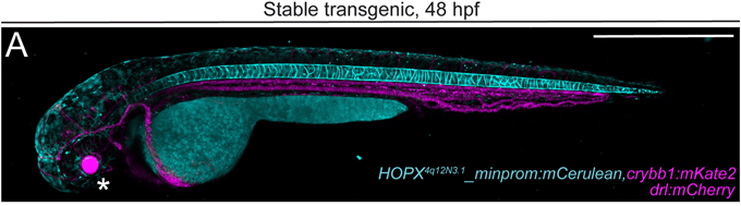
Figure 1: A stable transgenic line showing mCerulean expression in the vacuolar and sheath cells of the zebrafish notochord, mKate2 expression in the eye lens (asterisk), and mCherry expression in the cardiovascular system and blood at 2 days post-fertilization. Adapted from Kemmler et al., 2023, under a CC BY 4.0 license. |
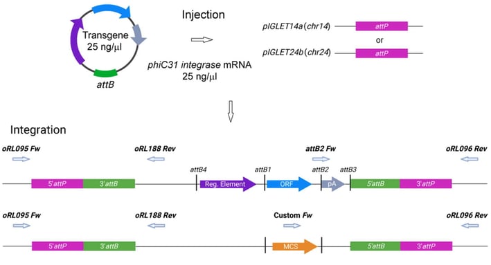
What if random integration won’t cut it? The Mosimann lab has also developed pIGLET, or phiC31 Integrase Genomic Loci Engineered for Transgenesis. Using a protocol that can be adapted to any Tol2 transgene site, they used CRISPR to insert landing sites for the phiC31 integrase at safe harbor loci they characterized. The vectors they provide can generate expression plasmids with various reporters for use in animals modified with these landing sites.
 |
|
Figure 2: Schematic of pIGLET system workflow. Transgenes with attB-containing pDEST or multiple cloning site (MCS) backbones are coinjected with phiC31 integrase mRNA into pIGLET14a or pIGLET24b zebrafish. Integration is depicted with both attB-containing pDEST (pCM268, pRL055, pRL056, pCK122, and pCK123) (top) or MCS (pRL092 and pRL093) (bottom) backbones. Adapted from Lalonde et al., 2024, under a CC BY-NC license. |
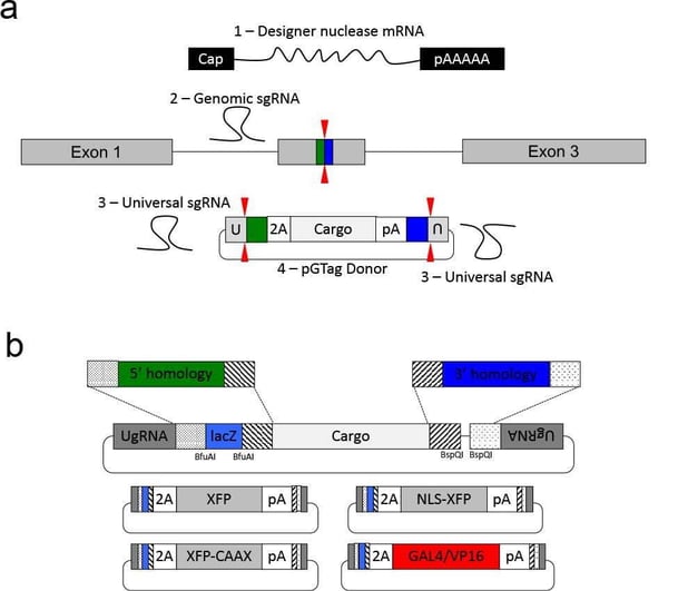
Looking for reporter gene knock-ins? The Jeffrey Essner lab has you covered. Their approach uses short homology arms to direct precise integrations that preserve transcription reading frame. While their vector design relies on CRISPR, the genome cutting step is compatible with CRISPR or TALENs. They provide donor vectors with red or green fluorescence and optional nuclear or membrane localization sequences, ready for you to clone in homology arms designed with their online tool.
 |
|
Figure 3: GeneWeld strategy and pGTag vector series. (A) GeneWeld reagent components are designed for simultaneous nuclease targeting of genome and donor to reveal short regions of homology. Red arrowheads represent nuclease DSB cut sites. Components include: 1 - Designer nuclease mRNA, either Cas9 to target both the genome and donor, or Cas9 to target the donor and TALEN to cut the genome; 2 - sgRNA for targeting Cas9 to the genome; 3 - Universal sgRNA to liberate donor cargo and homologous ends; and 4 - pGTag donor of interest with short homology arms. (B) Stippled and striped boxes represent sticky ends created by Type IIs restriction endonucleases BfuAI and BspQI, allowing digestion and ligation of both homology arms into the donor vector in a single reaction. Homology arm fragments are formed by annealing complementary oligonucleotides to form dsDNA with sticky ends for directional cloning into the vector. XFP = Green or Red Fluorescent Protein. pA = SV40 or β-actin 3’ untranslated region. NLS = nuclear localization sequence. CAAX = membrane localization sequence. Adapted from Wierson et al., 2020, under a CC BY 4.0 license. |
CRISPR/Cas9 genome editing
These famous bacterial scissors have made their way to zebrafish. Cut and paste, erase and replace — whatever metaphor you prefer, there’s probably a gene editing tool that does it.
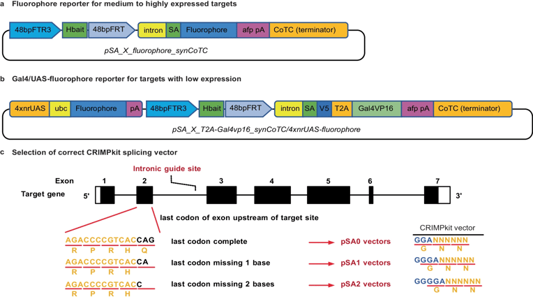
CRIMPkit is a no-assembly-required tool deposited by the Robert Bryson-Richardson lab. It relies on non-homologous end joining to insert a reporter into an intron of a target gene, interrupting its expression and allowing study of its native promoter. Since the kit includes variants for each open reading frame — plus is already codon optimized for zebrafish — CRIMPkit requires no customization to get started with insertional mutagenesis at your gene of interest.
 |
|
Figure 4: CRIMPkit vectors contain a guide RNA target site (Hbait) for CRISPR/Cas9 mediated linearization. The Hbait site is directly upstream of the 3’ region of β-actin intron 2, which contains the splice acceptor for exon 3. One or two bases have been added following the splice acceptor in pSA1 and pSA2 vectors respectively, to ensure the downstream reading frame is preserved. CRIMP vectors with a fluorophore after the splice acceptor (A) are designed for targeting genes with high expression levels. Vectors with Gal4vp16 after the splice acceptor and downstream UAS:fluorophore cassette (B) are for targeting genes with low expression levels. SA = splice acceptor, afp pA = Ocean pout anti-freeze protein 3’UTR until the poly-A sequence, ubc = 5’UTR intron of ubiquitin C CoTC co-transcriptional cleavage. (C) Selection of the correct CRIMP splicing vector. The reading frame of the last codon in the exon upstream of the intronic target site determines the correct vector to use in order to preserve the correct reading frame of the downstream coding sequence in a successfully targeted intron. Black bases indicate the final codon of an exon upstream of an intronic target. Blue bases indicate the sequence incorporated in CRIMP vectors to maintain the reading frame after splicing. Adapted from Miles et al., 2024, under a CC-BY 4.0 license. |
For those of you looking only to tweak a gene instead of disrupt it, the Gaurav Varshney lab has optimized adenine base editing for zebrafish. Their suite of ABE-ultramax editors includes variants with different targeting windows; an SpRY near-PAMless option; and a nanos promoter for exclusive germline expression, which allows lethal germline editing of progeny without harming the parent animal.
 |
|
Figure 5: A schematic diagram illustrates the editing window for ABE-Umax tools. Editing preferences are indicated by a red shading gradient, with the darkest red marking the most preferred position and lighter shades representing less preferred edits. A blue line highlights the precise editing position. The red triangle denotes the theoretical SpCas9 cutting site, while the PAM sequence and its complement are shown in green. Reproduced from Qin et al., 2024, under a CC-BY 4.0 license. |
Genome editing is great and all, but what about transcriptome editing? The Miguel Angel Moreno-Mateos lab has optimized CRISPR-Cas RNA endonucleases for use in zebrafish, including improving nuclear targeting, reducing collateral cleavage, and addressing gRNA-specific toxicity issues. Variants Cas7-11 and DjCas13d are especially recommended for their lower off-target effects in zebrafish embryos than the previously described RfxCas13d.
Plus, one CRISPR alternative…
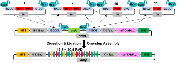
Mitochondrial DNA is stubbornly resistant to CRISPR editing, because RNA doesn’t cross the mitochondrial membrane. But have no fear! Transcription activator-like effectors (TALEs) are an effective alternative. These proteins use a pattern of repeat variable di-residues (RVDs) that each correspond to a DNA base, so assembly of these repeats can specify a DNA target sequence — no RNA required. The Bin Shen lab adapted a previously described TALE approach for cytosine base editing of mtDNA (Mok et al., 2020) to produce the DdCBE assembly kit for zebrafish, and demonstrated its use in modeling heritable mitochondrial diseases in these animals.
 |
|
Figure 6: Schematic overview of one-step assembly of DdCBE. Top, plasmids containing intended RVD are cleaved using BsaI to produce sticky ends. Middle, the RVDs with sticky ends are allowed to ligate to backbones sequentially. Bottom, the assembled DdCBE vectors contain 13.5–20.5 RVDs. 27 amino acids (aa) are put after ccdb in the backbone, and 14 aa are put in half RVD module. There are a total of 41 aa at C-terminus of TALE after assembly. Reproduced from Guo et al., 2021, under a CC-BY 4.0 license. |
Fluorescent proteins, biosensors, and optogenetics
That famous zebrafish transparency really lends itself to these glowing reporters!
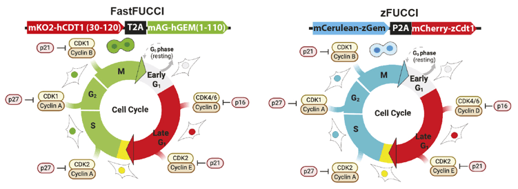
The Joshua Wythe lab offers an entire MAGIC toolbox of cloning vectors — Multisite Assembly of Gateway Induced Clones, that is. You can check out all 161 plasmids; we’ll just highlight the FUCCI cell cycle reporters (Sakaue-Sawano et al., 2008), which involve expressing fluorescent reporters tagged with opposite markers for cell cycle-dependent degradation. The team put FUCCI reporter variants into middle Gateway clones to be mixed and matched with promoters, reporters, regulation methods, and destination vectors from the same kit.
 |
|
Figure 7: Labeling cell cycle dynamics using FUCCI. Schematic of the FastFUCCI system (left, Koh et al., 2017) and the zFUCCI system (right, Bouldin et al., 2014). Reproduced from Gillespie et al., 2025, under a CC-BY 4.0 license. |
If you’re looking for calcium sensors, Addgene’s collection includes a variety of choices. The Eric Schreiter lab built a near-infrared HaloTag calcium indicator around a dye-ligand already known to cross the blood-brain barrier. Other colors are also compatible, including several that reach the central nervous system in living animals. The sensor is quenched by tryptophan in the absence of calcium, leading to the name WHaloCaMP: W (the abbreviation for tryptophan) + HaloTag + Ca2+-modulated protein (CaMP).
 |
|
Figure 8: Schematic representation of WHaloCaMP, showing domain arrangement, covalent binding of the dye-ligand, and the quenching tryptophan. Reproduced from Farrants et al., 2024, under a CC-BY 4.0 license. |
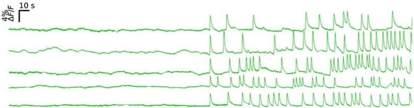
Ba-dum, ba-dum… The Adam Cohen lab developed and shared calcium and voltage sensors expressed specifically in the developing zebrafish heart. These tools let them watch these cells fire to produce the embryo’s first heartbeat.
 |
|
Figure 9: Multiplexed time-lapse calcium imaging captures the first heartbeats of zebrafish embryos. jGCaMP7f ΔF/F measurements of representative hearts aligned by the first calcium spike show abrupt onset of cardiac activity. Reproduced from Jia et al., 2022, under a CC-BY-NC-ND 4.0 license. |
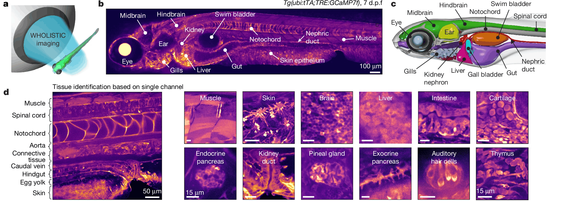
Tissue-specific expression just not cutting it? The Chie Satou lab developed WHOLISTIC, or WHole-Organism Live-Imaging System for recording Tissue and IntraCellular activity. By pairing their data collection and analysis workflow (which accounts for motion in a living animal) with whole-body expression of a calcium sensor, they observed signaling that stretched across tissues and new functional subdivisions of well-studied muscles.
 |
|
Figure 10: (A) Schematic of WHOLISTIC. (B) Transgenic zebrafish (7 days post-fertilization) expressing GCaMP7f pancellularly, imaged with spinning-disk confocal microscopy; samples were mounted laterally for visceral access. (C) Annotated 3D anatomical model of young zebrafish derived from whole-body expansion microscopy (WB-ExM) data. (D) Single fluorescence channel enables identification of organs and tissues via distinct visual textures (enlarged examples). Reproduced from Ruetten et al., 2026, under a CC-BY 4.0 license. |
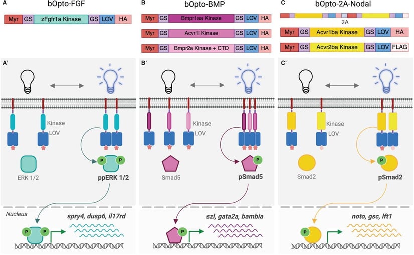
For tools that are activated by light instead of emitting it, check out the Katherine Rogers lab’s optogenetic toolset. They validated tools for light-activated expression of bone morphogenetic protein (BMP), fibroblast growth factor (FGF), and Nodal, three key signaling molecules in early vertebrate development. In each case, proteins that dimerize in response to light were coupled to zebrafish signaling kinases, which are activated by proximity. The Opto-FGF construct is also available in a Tol2 transgenesis-compatible format.
 |
|
Figure 11: Optogenetic signaling activator toolkit. (A,B,C) Schematic of constructs used to activate FGF (A), BMP (B), and Nodal (C) signaling. Myr = myristoylation motif, GS = glycine/serine linker, LOV = light oxygen voltage-sensing domain, HA = hemagglutinin tag, FLAG = FLAG epitope, P = phosphate group. The single-transcript bOpto-2A-Nodal construct (C) follows the same design as -FGF and -BMP, except the type I (Acvr1ba) and type II (Acvr2ba) components are connected via a 2A peptide sequence (gray). (A’,B’,C’) Optogenetic strategy to activate FGF (A’), BMP (B’), and Nodal (C’) signaling. Blue light-dimerizing LOV domains are fused to myristoylated receptor kinase domains. Blue light exposure should lead to receptor kinase interactions, signaling effector phosphorylation and activation of target genes. Reproduced from Iannucci et al., 2025, under a CC0 license. |
Lineage tracing and more
Genealogy isn’t just for history buffs — individual cells have unique lineages too, and they’re especially interesting early in development and during regenerative healing. But how to tell these cells apart?
The Alex Schier lab originally brought Brainbow to zebrafish. This colorful approach uses three fluorescent proteins whose combinations produce a rainbow of colors. The three colors are each flanked by two incompatible loxP sites, so random Cre recombination results in different color outcomes for different cells. Zebrabow places this construct under the control of a ubi promoter for ubiquitous zebrafish expression or a UAS enhancer for Gal4-inducible expression.
Another adaptation of this rainbow system comes from the Florence Marlow lab. Their team developed lineage tracing systems for germ cells specifically: Germ traCre combines germline-specific mCherry expression silenced by a floxed stop cassette with a photoactivatable Cre, expressed from an injected mRNA with a germ-cell-specific 3′ UTR. The result is mosaic labeling specific to germ cells. For a multicolor approach, Germbow brings the Brainbow/Zebrabow construct to this germline system. The lab used these constructs to trace primordial germ cell contributions to the adult gonad.
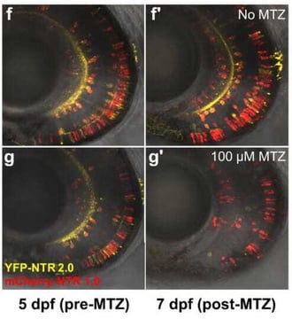
Among the zebrafish’s admirable qualities is its ability to regenerate, even from serious injuries such as those to the spinal cord. Studying this process requires targeted cell ablation, like with nitroreductases (NTR), which can convert prodrugs into toxic products and selectively kill cells that express them. The Jeff Mumm lab developed an improved NTR system, NTR 2.0, by engineering an alternative NTR enzyme to function efficiently at 100-fold lower doses of prodrug. That means fewer side effects and potentially longer treatment periods. Their approach works in resistant cell types and across bacteria, mammalian cell culture, and (of course) zebrafish, and their plasmids come with a choice of transgenic markers and expression systems.
 |
|
Figure 12: Ablation efficacy comparison of NTR 1.0 and NTR 2.0. Triple transgenic zebrafish larvae expressing YFP and NTR 2.0 (yellow cells) and/or mCherry and NTR 1.0 (red cells) were exposed to 100 μM MTZ [prodrug] for 48 h from 5–7 days post fertilization (dpf). Adapted from Sharrock et al., 2020, under a CC-BY-NC 4.0 license. |
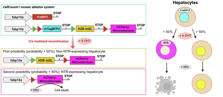
Once you’ve decided on targeted ablation, CellCousin2 can help you monitor which cells aid in the recovery. The Sumeet Pal Singh lab’s CellCousin system uses an inducible Cre to drive recombination of a cell-type-specific reporter construct. Unrecombined cells express blue fluorescent protein; after recombination, cells either express mGreenLantern or an mCherry-NTR fusion. Upon prodrug treatment, only green cells are left; their daughters inherit green fluorescence, while newly differentiated, unrecombined cells begin fluorescing blue. In the CellCousin2 improvements, the team offers several options to reduce background recombination and an option to use the NTR 2.0 enzyme described above.
 |
|
Figure 13: A Schematic overview of the CellCousin zebrafish model, which enables inducible, mosaic expression of Nitroreductase (NTR) in hepatocytes for targeted ablation. Hepatocyte-specific expression is driven by the fabp10a promoter. In the absence of recombination, hepatocytes express nuclear-localized mTagBFP2. Upon 4-hydroxytamoxifen (4-OHT) induction, Cre-mediated recombination results in the stochastic expression of either H2B-mGL (nuclear GFP) or mCherry-NTR fusion protein. Following treatment with Metronidazole (Mtz), only mCherry-NTR–expressing hepatocytes undergo cell death, allowing for controlled, partial ablation within the liver. Reproduced from Hovhannisyan et al., 2026, under a CC BY-NC-ND 4.0 license. |
Conclusion
The best part of this round-up is seeing so many tools that are inter-operable and even build on one another. That’s what we love about open science! The labs that have shared their work, both those highlighted here and beyond, are the backbone (perhaps we should say notochord?) of the zebrafish science community. Whether you mix, match, or make your own, major thanks to the depositors and users who have made Addgene’s zebrafish collection strong!
Explore Zebrafish Plasmids at Addgene
References and Resources
References
Cloning and transgenesis
Don, E. K., Formella, I., Badrock, A. P., Hall, T. E., Morsch, M., Hortle, E., Hogan, A., Chow, S., Gwee, S. S. L., Stoddart, J. J., Nicholson, G., Chung, R., & Cole, N. J. (2017). A Tol2 Gateway-Compatible Toolbox for the Study of the Nervous System and Neurodegenerative Disease. Zebrafish, 14(1), 69–72. https://doi.org/10.1089/zeb.2016.1321
Kemmler, C. L., Moran, H. R., Murray, B. F., Scoresby, A., Klem, J. R., Eckert, R. L., Lepovsky, E., Bertho, S., Nieuwenhuize, S., Burger, S., D’Agati, G., Betz, C., Puller, A.-C., Felker, A., Ditrychova, K., Bötschi, S., Affolter, M., Rohner, N., Lovely, C. B., … Mosimann, C. (2023). Next-generation plasmids for transgenesis in zebrafish and beyond. Development, 150(8), dev201531. https://doi.org/10.1242/dev.201531
Lalonde, R. L., Wells, H. H., Kemmler, C. L., Nieuwenhuize, S., Lerma, R., Burger, A., & Mosimann, C. (2024). pIGLET: Safe harbor landing sites for reproducible and efficient transgenesis in zebrafish. Science Advances, 10(23), eadn6603. https://doi.org/10.1126/sciadv.adn6603
Wierson, W. A., Welker, J. M., Almeida, M. P., Mann, C. M., Webster, D. A., Torrie, M. E., Weiss, T. J., Kambakam, S., Vollbrecht, M. K., Lan, M., McKeighan, K. C., Levey, J., Ming, Z., Wehmeier, A., Mikelson, C. S., Haltom, J. A., Kwan, K. M., Chien, C.-B., Balciunas, D., … Essner, J. (2020, May 15). Efficient targeted integration directed by short homology in zebrafish and mammalian cells. eLife Sciences Publications Limited. eLife. https://doi.org/10.7554/eLife.53968
CRISPR/Cas9 genome editing
Miles, L. B., Calcinotto, V., Oveissi, S., Serrano, R. J., Sonntag, C., Mulia, O., Lee, C., & Bryson-Richardson, R. J. (2024). CRIMP: A CRISPR/Cas9 insertional mutagenesis protocol and toolkit. Nature Communications, 15(1), 5011. https://doi.org/10.1038/s41467-024-49341-7
Qin, W., Liang, F., Lin, S.-J., Petree, C., Huang, K., Zhang, Y., Li, L., Varshney, P., Mourrain, P., Liu, Y., & Varshney, G. K. (2024). ABE-ultramax for high-efficiency biallelic adenine base editing in zebrafish. Nature Communications, 15(1), 5613. https://doi.org/10.1038/s41467-024-49943-1
Moreno-Sánchez, I., Hernández-Huertas, L., Nahón-Cano, D., Martínez-García, P. M., Treichel, A. J., Gómez-Marin, C., Tomás-Gallardo, L., da Silva Pescador, G., Kushawah, G., Egidy, R., Perera, A., Díaz-Moscoso, A., Cano-Ruiz, A., Walker, J. A., Muñoz, M. J., Holden, K., Galcerán, J., Nieto, M. Á., Bazzini, A. A., & Moreno-Mateos, M. A. (2025). Enhanced RNA-targeting CRISPR-Cas technology in zebrafish. Nature Communications, 16(1), 2591. https://doi.org/10.1038/s41467-025-57792-9
One CRISPR alternative
Guo, J., Zhang, X., Chen, X., Sun, H., Dai, Y., Wang, J., Qian, X., Tan, L., Lou, X., & Shen, B. (2021). Precision modeling of mitochondrial diseases in zebrafish via DdCBE-mediated mtDNA base editing. Cell Discovery, 7(1), 78. https://doi.org/10.1038/s41421-021-00307-9
Mok, B. Y., de Moraes, M. H., Zeng, J., Bosch, D. E., Kotrys, A. V., Raguram, A., Hsu, F., Radey, M. C., Peterson, S. B., Mootha, V. K., Mougous, J. D., & Liu, D. R. (2020). A bacterial cytidine deaminase toxin enables CRISPR-free mitochondrial base editing. Nature, 583(7817), 631–637. https://doi.org/10.1038/s41586-020-2477-4
Fluorescent proteins, biosensors, and optogenetics
Anderson, W. K., Iannucci, L. E., Sinaii, N., Porcino, J., & Rogers, K. W. (2026). A transgenic zebrafish for spatiotemporal optogenetic activation of FGF/ERK signaling. Biology Open, 15(9), bio062695. https://doi.org/10.1242/bio.062695
Bouldin, C. M., Snelson, C. D., Farr, G. H., & Kimelman, D. (2014). Restricted expression of cdc25a in the tailbud is essential for formation of the zebrafish posterior body. Genes & Development, 28(4), 384–395. https://doi.org/10.1101/gad.233577.113
Farrants, H., Shuai, Y., Lemon, W. C., Monroy Hernandez, C., Zhang, D., Yang, S., Patel, R., Qiao, G., Frei, M. S., Plutkis, S. E., Grimm, J. B., Hanson, T. L., Tomaska, F., Turner, G. C., Stringer, C., Keller, P. J., Beyene, A. G., Chen, Y., Liang, Y., … Schreiter, E. R. (2024). A modular chemigenetic calcium indicator for multiplexed in vivo functional imaging. Nature Methods, 21(10), 1916–1925. https://doi.org/10.1038/s41592-024-02411-6
Gillespie, W. B., Zhang, Y., Ruiz, O. E., Cerda, J., Ortiz-Guzman, J., Sherman, M., Turner, W. D., Largoza, G., Mosser, L. E., Fujimoto, E., Chien, C.-B., Kwan, K. M., Arenkiel, B. R., Devine, W. P., & Wythe, J. D. (2025). MultiSite Assembly of Gateway Induced Clones (MAGIC): A flexible cloning toolbox for use in vertebrate model systems. Development, 152(22), dev204308. https://doi.org/10.1242/dev.204308
Iannucci, L. E., Maria Thomas, V. S., Anderson, W. K., Murphy, M. R., Donahue, C. E. T., Campbell, C. E., Monaghan, M. T., Saul, A. J., & Rogers, K. W. (2026). An optogenetic toolkit for robust activation of FGF, BMP, & Nodal signaling in zebrafish. Developmental Biology, 536, 155–171. https://doi.org/10.1016/j.ydbio.2026.05.003
Iannucci, L. E., Thomas, V. S. M., Murphy, M. R., Donahue, C. E. T., Anderson, W. K., Rogers, C. E., Saul, A. J., & Rogers, K. W. (2025). An optogenetic toolkit for robust activation of FGF, BMP, & Nodal signaling in zebrafish (2025.04.17.649426). bioRxiv: The Preprint Server for Biology. https://doi.org/10.1101/2025.04.17.649426
Jia, B. Z., Qi, Y., Wong-Campos, J. D., Megason, S. G., & Cohen, A. E. (2022). A bioelectrical phase transition patterns the first beats of a vertebrate heart (p. 2022.12.06.519309). bioRxiv. https://doi.org/10.1101/2022.12.06.519309
Jia, B. Z., Qi, Y., Wong-Campos, J. D., Megason, S. G., & Cohen, A. E. (2023). A bioelectrical phase transition patterns the first vertebrate heartbeats. Nature, 622(7981), 149–155. https://doi.org/10.1038/s41586-023-06561-z
Koh, S.-B., Mascalchi, P., Rodriguez, E., Lin, Y., Jodrell, D. I., Richards, F. M., & Lyons, S. K. (2017). A quantitative FastFUCCI assay defines cell cycle dynamics at a single-cell level. Journal of Cell Science, 130(2), 512–520. https://doi.org/10.1242/jcs.195164
Rogers, K. W., ElGamacy, M., Jordan, B. M., & Müller, P. (2020). Optogenetic investigation of BMP target gene expression diversity. eLife, 9, e58641. https://doi.org/10.7554/eLife.58641
Ruetten, V. M. S., Zheng, W., Siwanowicz, I., Mensh, B. D., Eddison, M., Hu, A., Chi, Y., Lemire, A. L., Guo, C., Kadobianskyi, M., Renz, M., Lelek-Greskovic, S., He, Y., Close, K., Ihrke, G., Dev, A., Petruncio, A., Wan, Y., Zhang, R., … Ahrens, M. B. (2026). Imaging cellular activity across all organs reveals body-wide circuits. Nature, 1–11. https://doi.org/10.1038/s41586-026-10979-6
Lineage tracing and more
Pahima, M., & Marlow, F. L. (2026). Primordial germ cells differentially contribute to the germline in zebrafish. Development, 153(15), dev204904. https://doi.org/10.1242/dev.204904
Pan, Y. A., Freundlich, T., Weissman, T. A., Schoppik, D., Wang, X. C., Zimmerman, S., Ciruna, B., Sanes, J. R., Lichtman, J. W., & Schier, A. F. (2013). Zebrabow: Multispectral cell labeling for cell tracing and lineage analysis in zebrafish. Development, 140(13), 2835–2846. https://doi.org/10.1242/dev.094631
Hovhannisyan, G. G., Akhourbi, T., Eski, S. E., Pirson, I., Gurzov, E. N., & Singh, S. P. (2026). CellCousin2: An optimized system for partial ablation and tracing of regenerative lineages. NPJ Regenerative Medicine, 11(1), 23. https://doi.org/10.1038/s41536-026-00473-y
Sharrock, A. V., Mulligan, T. S., Hall, K. R., Williams, E. M., White, D. T., Zhang, L., Emmerich, K., Matthews, F., Nimmagadda, S., Washington, S., Le, K. D., Meir-Levi, D., Cox, O. L., Saxena, M. T., Calof, A. L., Lopez-Burks, M. E., Lander, A. D., Ding, D., Ji, H., … Mumm, J. S. (2022). NTR 2.0: A rationally engineered prodrug-converting enzyme with substantially enhanced efficacy for targeted cell ablation. Nature Methods, 19(2), 205–215. https://doi.org/10.1038/s41592-021-01364-4
Sharrock, A. V., Mulligan, T. S., Hall, K. R., Williams, E. M., White, D. T., Zhang, L., Matthews, F., Nimmagadda, S., Washington, S., Le, K., Meir-Levi, D., Cox, O. L., Saxena, M. T., Calof, A. L., Lopez-Burks, M. E., Lander, A. D., Ding, D., Ji, H., Ackerley, D. F., & Mumm, J. S. (2020). NTR 2.0: A rationally-engineered prodrug converting enzyme with substantially enhanced efficacy for targeted cell ablation (p. 2020.05.22.111427). bioRxiv. https://doi.org/10.1101/2020.05.22.111427
Additional resources on the Addgene blog
- Plasmids 101: Gateway Cloning
- Plasmids 101: Cre-lox
- CRISPR 101: Non-Homologous End Joining
- CRISPR 101: Cytosine and Adenine Base Editors
Resources on addgene.org
Topics: Model Organisms



Leave a Comment