By Guest Blogger
Read More
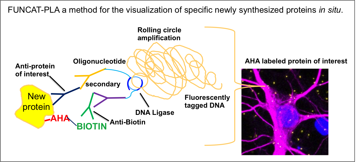
This post was contributed by guest blogger, Eugenia Rojas. A question worthy of a PhD: How do you visualize protein turnover within a neuron? For my PhD I studied a synaptic protein that is linked to neurodegeneration. The level of this protein is decreased in Alzheimer’s ...
Since the discovery of GFP over 50 years ago, the growing spectrum of fluorescent proteins (FPs) has been an invaluable resource for studying the organization and function of cellular systems. FPs have been used to track protein localization, cell structure, intracellular ...
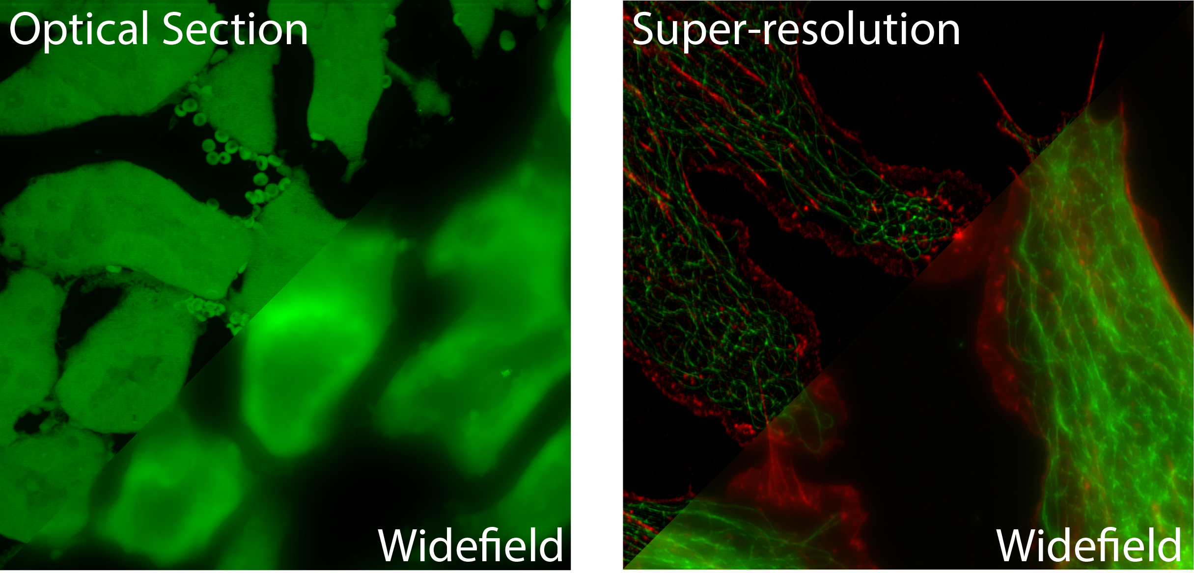
This post was contributed by Doug Richardson, Director of the Harvard Center for Biological Imaging and a Lecturer on Molecular and Cellular Biology at Harvard University. No matter whether you are a sports photographer at the Super Bowl, a medical technologist taking an x-ray, ...
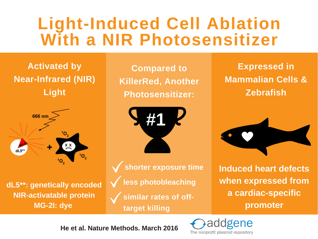
Have you ever wanted to selectively kill a subset of cells in your model system? Turns out that with light-inducible photosensitizers and a quick zap of the proper color light, you can do just that. Photosensitizing dyes and proteins have been around for awhile (check out this ...
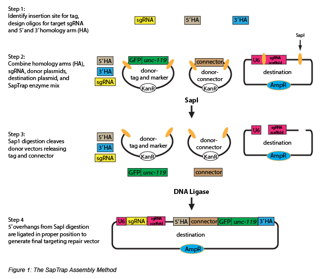
In complex metazoans, rapid cell division and large scale cell mobility are essential processes during embryonic development. These are required for a growing organism to make the complicated transition from a clump of cells to a fully differentiated body. In contrast, these ...
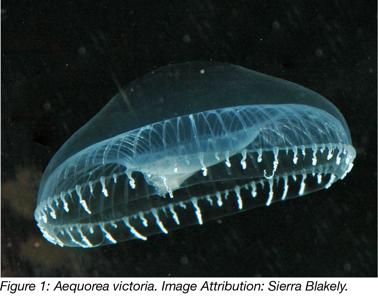
Luminescent molecules are very useful tools because we can easily detect and measure the light they emit. Proteins that give off light include chemiluminescent proteins, like luciferases, as well as fluorescent ones, like Green Fluorescent Protein (GFP). These molecules occur ...
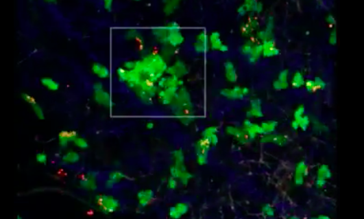
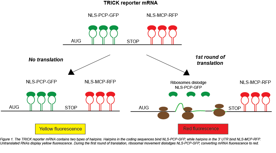
Regulating translation is key to cellular function, especially during development or stress. With ribosome profiling, researchers have been able to study the effects of various stimuli on global translation, but a visual technique to study translation remained elusive. Two ...